Accession No
0196
Brief Description
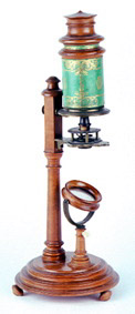
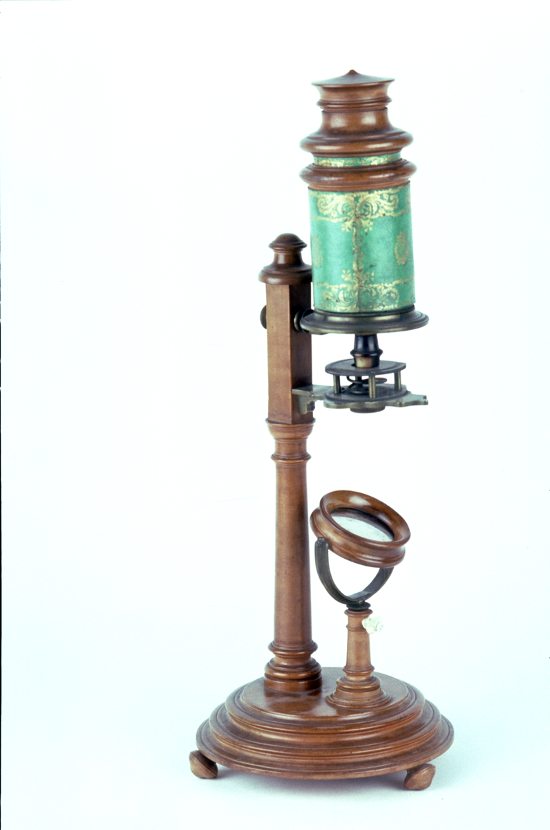
compound microscope, side pillar type, by Johann Balthasar Oppelt, German, 1786 - 1790
Origin
Germany; Ansbach
Maker
Oppelt, Johann Balthasar
Class
microscopes
Earliest Date
1786
Latest Date
1790
Inscription Date
Material
wood ([pear-wood], lignum vitae); glass; paper (pasteboard); hide (leather); metal (brass); organic (horn)
Dimensions
height 470mm; base diameter 180mm
Special Collection
Robert Whipple collection
Provenance
Purchased by Robert Stewart Whipple at an auction. This object was part of the Crisp Collection, and was sold as Lot 124 at the auction of this collection held on 17/02/1925 at the Steven’s Auction Rooms. The purchase price included a commission to T.H. Court. A photocopy of the Crisp inventory record is contained in the history file, and records that the object was “Found in the attic.”
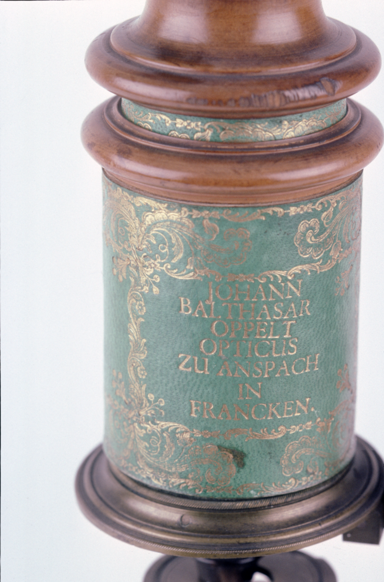
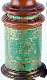
Inscription
JOHANN / BALTHASAR / OPPELT / OPTICUS / ZU ANSPACH / IN FRANKEN
Description Notes
Turned [pearwood] base with 3 knob feet. Swinging mirror on pear-wood stand with ivory clamp. Turned [pearwood] pillar with brass stage plate and spring stage (later screw fixing). Pasteboard collar covered with green, tooled leather with wooden ferrule either end and brass ferrule for attachment to pillar. Pasteboard body covered in green tooled leather with pear-wood snout and screw fit objective in a horn cell. Field and eye lenses in a horn and lignum vitae cell, which screw fits into pear-wood collar. Pear-wood dust cap.
References
Events
Description
This type of microscope was first designed by Henry Baker, a microscopist, and John Cuff, an instrument maker, in 1743. By mounting the stage on a side pillar the instrument became easier to use, with the operating parts much more accessible than in previous designs. The focus was controlled by a finely tuned screwthread, and was thus made far more accurate.
More on compound microscopes
The compound microscope was developed during the 17th Century and was closely related to the refracting telescope. Its popularity increased after the publication in 1665 of Robert Hooke’s (1635-1703) Micrographia. Micrographia contained detailed pictures, never before seen, of insects magnified using a compound microscope.
A compound microscope uses two or more lenses. The lenses are held at certain distances from each other and are mounted inside a rigid tube. The tube was usually made from pasteboard, ivory, or most commonly, brass. The basic compound microscope magnifies an image in two stages -
Stage one: Light from a mirror is reflected up through the specimen into a powerful objective lens.
Stage two: The image produced by the objective lens is magnified again by the eye lens, which works like a simple magnifying lens.
The first compound microscope consisted of a simple barrel which would have been held up to the light. Later developments ensured that the compound microscope had a stable base, usually a brass stand and a side pillar.
In the 17th Century, the compound microscope had some serious drawbacks which made it easier to use a simple microscope (which have only one lens) instead. The image produced by a compound microscope was often affected by two types of aberration, known as chromatic and spherical. These aberrations caused blurring to the image (spherical) and the edge of the specimen to colour (chromatic). Chromatic aberration was removed at the end of the 18th Century by Harmanus van Deijlan, an instrument maker in Amsterdam. In 1830, spherical aberration was overcome by Joseph Lister who developed the achromatic lens. Achromatic lenses became widely used in microscopes in the 1850s and are still used today.
FM:42146
Images (Click to view full size):