Accession No
1003
Brief Description
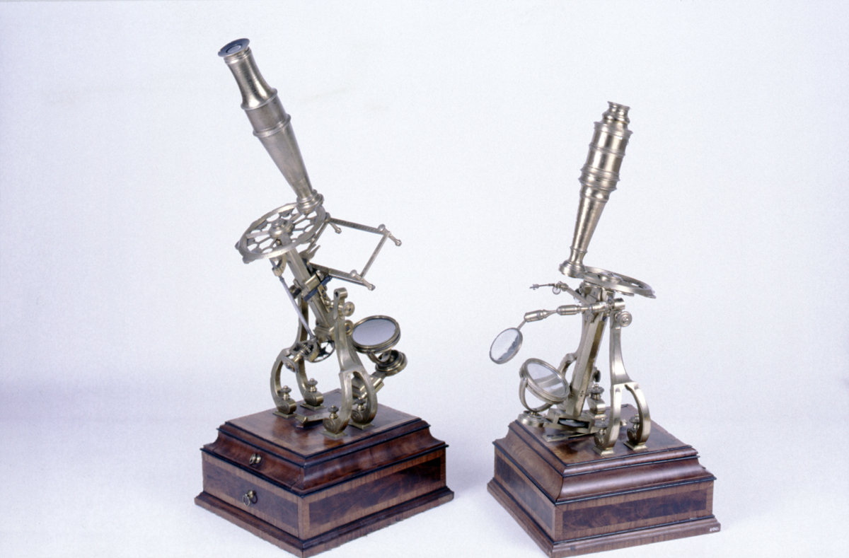
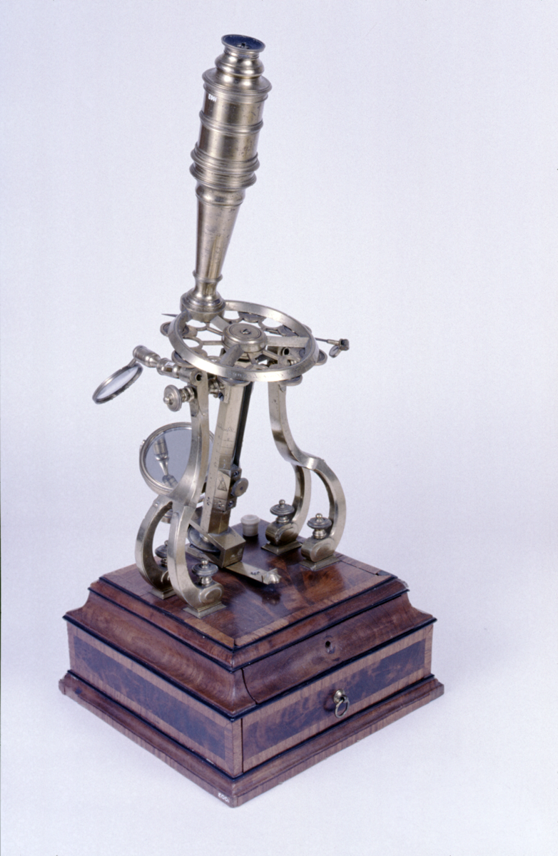
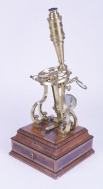
compound microscope, side pillar ‘prince of wales’ type, by George Adams Snr., English, circa 1760
Origin
England; London; Fleet Street
Maker
Adams, George (Snr.)
Class
microscopes
Earliest Date
1760
Latest Date
1760
Inscription Date
Material
wood (mahogany, satinwood?); metal (brass); glass; ivory
Dimensions
height 460mm; breadth 200mm; depth 200mm
Special Collection
Provenance
On loan from Gonville & Caius College, University of Cambridge from 1950.
Inscription
‘Made by G.Adams Fleet Street LONDON’
Description Notes
Mahogany box foot with [silk-wood or satinwood] veneer; 2 drawers in base; brass; 2 arched supports divided at base to form 4 feet with knurled screws fixing them to the base; limb pivots between the supports; limb fixed to box foot by a spring which allows the limb to tilt when released; limb an octagonal column with bar for swinging plano/concave mirror; body carried on rod inside column with sliding coarse focus marked 2-8 on pillar with knurled knob on plate with engraved hand motif; fine focus by knurled screw at the base of the column; dovetailed slide for stage fittings; fretwork wheel of objectives marked ‘1-8’; with screw thread for body; tapered snout screws to collar; screws to field lens collar; eye lens collar; eye shade with sliding dust cover. 1 stage single ring type with screw below; frog plate with dove tail slide; stage forceps with dove tail slide; condenser lens with articulated arm mounted on column; tweezers; talc box; lieberkuhn on holder which slides over the stage mount marked ‘single 4.5.6.’ and ‘Double 4.5.6.7.8.’, ivory [slide box]; second frog plate (cone and various other accessories in poor condition, [not original]).
References
Events
Description
The compound microscope was developed during the 17th Century and was closely related to the refracting telescope. Its popularity increased after the publication of Robert Hooke’s (1635-1703) Micrographia in 1665. Micrographia contained detailed pictures, never before seen, of insects magnified using a compound microscope.
A compound microscope uses two or more lenses. The lenses are held at certain distances from each other and are mounted inside a rigid tube. The tube was usually made from pasteboard, ivory, or most commonly, brass. The basic compound microscope magnifies an image in two stages -
Stage one: Light from a mirror is reflected up through the specimen into a powerful objective lens.
Stage two: The image produced by the objective lens is magnified again by the eye lens, which works like a simple magnifying lens.
The first compound microscope consisted of a simple barrel which would have been held up to the light. Later developments ensured that the compound microscope had a stable base, usually a brass stand and a side pillar.
In the 17th Century, the compound microscope had some serious drawbacks which made it easier to use a simple microscope (which have only one lens) instead. The image produced by a compound microscope was often affected by two types of aberrations known as chromatic and spherical. These aberrations caused blurring to the image (spherical) and the edge of the specimen to colour (chromatic). Chromatic aberration was removed at the end of the 18th Century by Harmanus van Deijlan, an instrument maker in Amsterdam. In 1830, spherical aberration was overcome by Joseph Lister, who developed the achromatic lens. Achromatic lenses became widely used in microscopes in the 1850s and are still used today.
From display label:
In addition to the amateur pursuits illustrated in this case, microscopy was a gentlemanly activity during the eighteenth century. This microscope is a somewhat reduced version of one made by Adams for the Prince of Wales.
FM:42175
Images (Click to view full size):