Accession No
3843
Brief Description
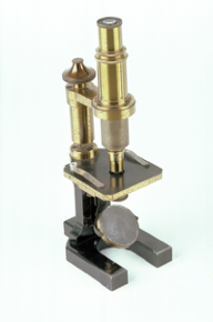
compound microscope, possibly achromatic, by Carl Zeiss, German, 2/2 19th Century
Origin
Germany; Jena
Maker
Carl Zeiss
Class
microscopes
Earliest Date
1850
Latest Date
1900
Inscription Date
Material
metal (brass, oxidised brass, white metal); glass; wood; cloth (velvet)
Dimensions
height 313mm; depth 132mm; breadth 90mm; box length 355mm; breadth 225mm; height 118mm
Special Collection
Provenance
Purchased from German seller, 04/1992.
Inscription
‘C. ZEISS
JENA
8285’ (limb brace)
‘1/4
R & J
BECK’ (objective)
‘C. ZEISS
AA
JENA’ (objective case)
‘CARL ZEISS
JENA
7x’ (eyepiece)
‘x5’ (other eyepiece)
Description Notes
Black-painted horseshoe base, flat profile. Flat pillar mounted on this with cylindrical upper section. Upper section of pillar, carrying limb and stage, pivots within lower section of pillar. Conical fine focus knurled knob at top of pillar. Plano-concave substage mirror on hinged arm with movement in all directions. Rectangular oxidised brass stage, with white metal substage clips. Horizontal bracket from pillar to limb. Coarse focus by movement of optical system within sleeve of limb. Draw tube divided [13.7] - 20, numbered by 1, subdivided to 0.1. Push-fit eyepiece; screw-fit objective.
Wooden box with white metal carrying handle, lock (no key), hook fasteners and hinges. Fitted and lined with blue velvet. Contains eyepiece and objective in brass screw-top case. Further spaces for lenses.
Condition fair (extensive corrosion on metal on outside of box); incomplete (lenses missing)
References
Events
Description
The solution to the problem of chromatic aberration
When light travels through an ordinary lens each colour is bent through a different angle. In a microscope this causes what is known as chromatic aberration, whereby a spectrum of colours will appear around the image being viewed. Chromatic aberration was a big drawback when using early versions of the compound microscope.
Chromatic aberration was overcome by Joseph Jackson Lister in 1830, after he developed the achromatic lens for microscopes. The new type of lens prevented colour separation by combining two lenses made of different types of glass. The first lens that light passed through would split the colours and the second lens acted to bring the colours back together again. This produced a much sharper and clearer image than it had previously been possible to achieve.
In the Victorian period the achromatic microscope became a vital tool in medical and scientific research. Improvements to the optical performance of the microscope saw developments in the design and construction of the microscope. The microscope became sturdier and could focus to a finer level.
Scientific instrument makers in the Victorian period who improved the rigidity of the optical tube and the focusing ability of the achromatic microscope include Andrew Ross, James Smith and Hugh Powell.
More on compound microscopes
The compound microscope was developed during the 17th Century and was closely related to the refracting telescope. Its popularity increased after the publication in 1665 of Robert Hooke’s (1635-1703) Micrographia. Micrographia contained detailed pictures, never before seen, of insects magnified using a compound microscope.
A compound microscope uses two or more lenses. The lenses are held at certain distances from each other and are mounted inside a rigid tube. The tube was usually made from pasteboard, ivory, or most commonly, brass. The basic compound microscope magnifies an image in two stages -
Stage one: Light from a mirror is reflected up through the specimen into a powerful objective lens.
Stage two: The image produced by the objective lens is magnified again by the eye lens, which works like a simple magnifying lens.
The first compound microscope consisted of a simple barrel which would have been held up to the light. Later developments ensured that the compound microscope had a stable base, usually a brass stand and a side pillar.
In the 17th Century, the compound microscope had some serious drawbacks which made it easier to use a simple microscope (which have only one lens) instead. The image produced by a compound microscope was often affected by two types of aberration, known as chromatic and spherical. These aberrations caused blurring to the image (spherical) and the edge of the specimen to colour (chromatic). Chromatic aberration was removed at the end of the 18th Century by Harmanus van Deijlan, an instrument maker in Amsterdam. In 1830, spherical aberration was overcome by Joseph Lister who developed the achromatic lens. Achromatic lenses became widely used in microscopes in the 1850s and are still used today.
FM:42176
Images (Click to view full size):