Accession No
3178
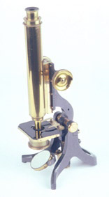
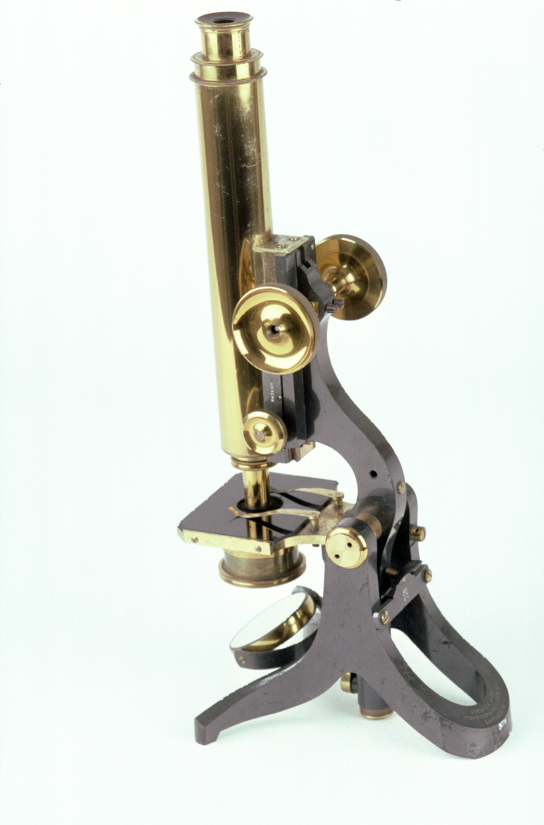
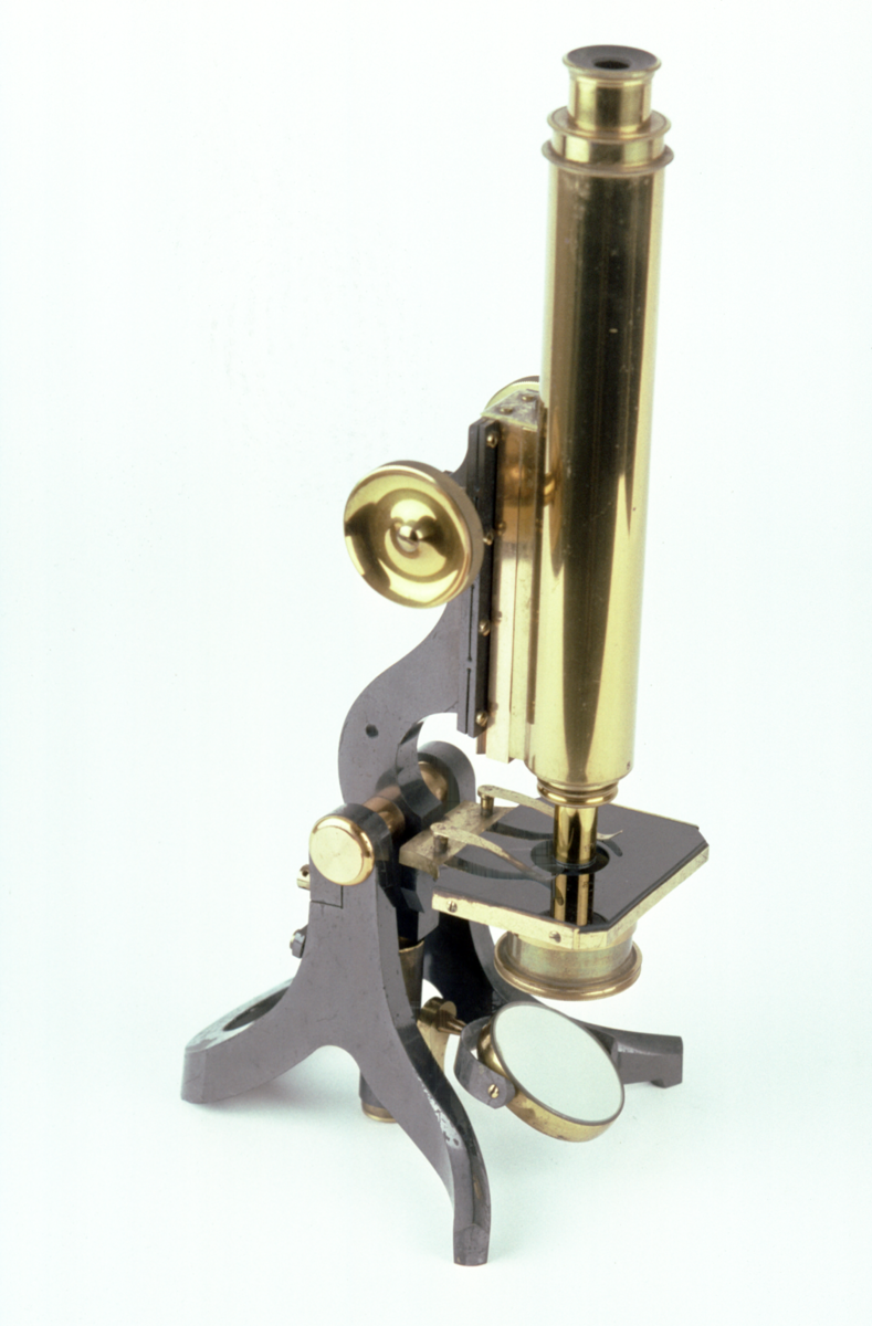
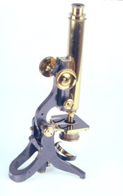
Brief Description
achromatic compound microscope, by Swift and Son, English, 1900 (c)
Origin
England; London; 81 Tottenham Court Road
Maker
Swift and Son
Class
microscopes
Earliest Date
1900
Latest Date
1900
Inscription Date
Material
metal (oxidised brass); glass
Dimensions
height 364mm; depth 167mm; breadth 143mm
Special Collection
Provenance
Belonged to the donor’s uncle, Cecil Warburton.
Inscription
‘SWIFT & SON
81 TOTTENHAM COURT RD
LONDON W.’
Trade mark on back of limb.
Description Notes
Horseshoe/kettledrum type foot in oxidised brass; double uprights to swinging bar supporting limb; tailpiece with swinging plano/concave mirror on sliding shoe; square stage with black glass ground and spring clips; substage condenser with central stop; arched limb with large knurled screw either side for coarse focus; [patented] clamping screw on side of limb; body with draw tube; push fit eyepiece; objective ‘1/6” SWIFT & SON’. Possibly ‘College No 15’ type microscope.
References
Events
Description
The solution to the problem of chromatic aberration
When light travels through an ordinary lens each colour is bent through a different angle. In a microscope this causes what is known as chromatic aberration, whereby a spectrum of colours will appear around the image being viewed. Chromatic aberration was a big drawback when using early versions of the compound microscope.
Chromatic aberration was overcome by Joseph Jackson Lister in 1830, after he developed the achromatic lens for microscopes. The new type of lens prevented colour separation by combining two lenses made of different types of glass. The first lens that light passed through would split the colours and the second lens acted to bring the colours back together again. This produced a much sharper and clearer image than it had previously been possible to achieve.
In the Victorian period the achromatic microscope became a vital tool in medical and scientific research. Improvements to the optical performance of the microscope saw developments in the design and construction of the microscope. The microscope became sturdier and could focus to a finer level.
Scientific instrument makers in the Victorian period who improved the rigidity of the optical tube and the focusing ability of the achromatic microscope include Andrew Ross, James Smith and Hugh Powell.
More on compound microscopes
The compound microscope was developed during the 17th Century and was closely related to the refracting telescope. Its popularity increased after the publication in 1665 of Robert Hooke’s (1635-1703) Micrographia. Micrographia contained detailed pictures, never before seen, of insects magnified using a compound microscope.
A compound microscope uses two or more lenses. The lenses are held at certain distances from each other and are mounted inside a rigid tube. The tube was usually made from pasteboard, ivory, or most commonly, brass. The basic compound microscope magnifies an image in two stages -
Stage one: Light from a mirror is reflected up through the specimen into a powerful objective lens.
Stage two: The image produced by the objective lens is magnified again by the eye lens, which works like a simple magnifying lens.
The first compound microscope consisted of a simple barrel which would have been held up to the light. Later developments ensured that the compound microscope had a stable base, usually a brass stand and a side pillar.
In the 17th Century, the compound microscope had some serious drawbacks which made it easier to use a simple microscope (which have only one lens) instead. The image produced by a compound microscope was often affected by two types of aberration, known as chromatic and spherical. These aberrations caused blurring to the image (spherical) and the edge of the specimen to colour (chromatic). Chromatic aberration was removed at the end of the 18th Century by Harmanus van Deijlan, an instrument maker in Amsterdam. In 1830, spherical aberration was overcome by Joseph Lister who developed the achromatic lens. Achromatic lenses became widely used in microscopes in the 1850s and are still used today.
FM:42933
Images (Click to view full size):