Accession No
0325
Brief Description
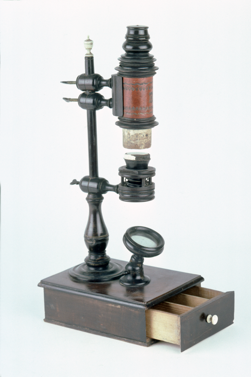
compound microscope, side pillar nuremburg ‘toy’ type, by J. F. F., German, 1/2 19th Century
Origin
Germany; Nuremburg
Maker
J. F. F.
Class
microscopes
Earliest Date
1800
Latest Date
1850
Inscription Date
Material
glass (mirror); metal (brass, other metal); paper; wood (mahogany, pine, pear)
Dimensions
height 459mm; depth 185mm; breadth 134mm; height to top of body 359mm
Special Collection
Robert Whipple collection
Provenance
Purchased from Gertrude Hamilton (trading as ’Mercator’), Paris, in 08/1926.
Inscription
Description Notes
Rectangular base with pine core and mahogany veneer; turned pear wood pillar with ivory vase finial; 3 side arms clamped by brass wing nuts; lowest to spring stage; upper to a clamp round body tube; body of pasteboard covered with imitation vellum with gold stamps; 2 draw-tubes covered with decorated paper each with a pear wood ferrule; conical snout with field lens and screw fit objective; eyepiece with dust cover; swinging mirror on pivoted base; 3 objectives. Maker’s mark “J.F.F.”.
References
Events
Description
The town of Nuremberg in Bavaria has long been famous for its production of finely constructed wooden toys and craft items. Among these were microscopes made from soft wood, with draw-tubes of card and patterned paper. A large volume of such items were made during the first half of the nineteenth century.
More on compound microscopes
The compound microscope was developed during the 17th Century and was closely related to the refracting telescope. Its popularity increased after the publication in 1665 of Robert Hooke’s (1635-1703 Micrographia. Micrographia contained detailed pictures, never before seen, of insects magnified using a compound microscope.
A compound microscope uses two or more lenses. The lenses are held at certain distances from each other and are mounted inside a rigid tube. The tube was usually made from pasteboard, ivory, or most commonly, brass. The basic compound microscope magnifies an image in two stages -
Stage one: Light from a mirror is reflected up through the specimen into a powerful objective lens.
Stage two: The image produced by the objective lens is magnified again by the eye lens, which works like a simple magnifying lens.
The first compound microscope consisted of a simple barrel which would have been held up to the light. Later developments ensured that the compound microscope had a stable base, usually a brass stand and a side pillar.
In the 17th Century, the compound microscope had some serious drawbacks which made it easier to use a simple microscope (which have only one lens) instead. The image produced by a compound microscope was often affected by two types of aberrations known as chromatic and spherical. These aberrations caused blurring to the image (spherical) and the edge of the specimen to colour (chromatic). Chromatic aberration was removed at the end of the 18th Century by Harmanus van Deijlan, an instrument maker in Amsterdam. In 1830, spherical aberration was overcome by Joseph Lister, who developed the achromatic lens. Achromatic lenses became widely used in microscopes in the 1850s and are still used today.
Created by: Corrina Bower
FM:42941
Images (Click to view full size):