Accession No
0009
Brief Description
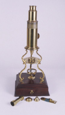
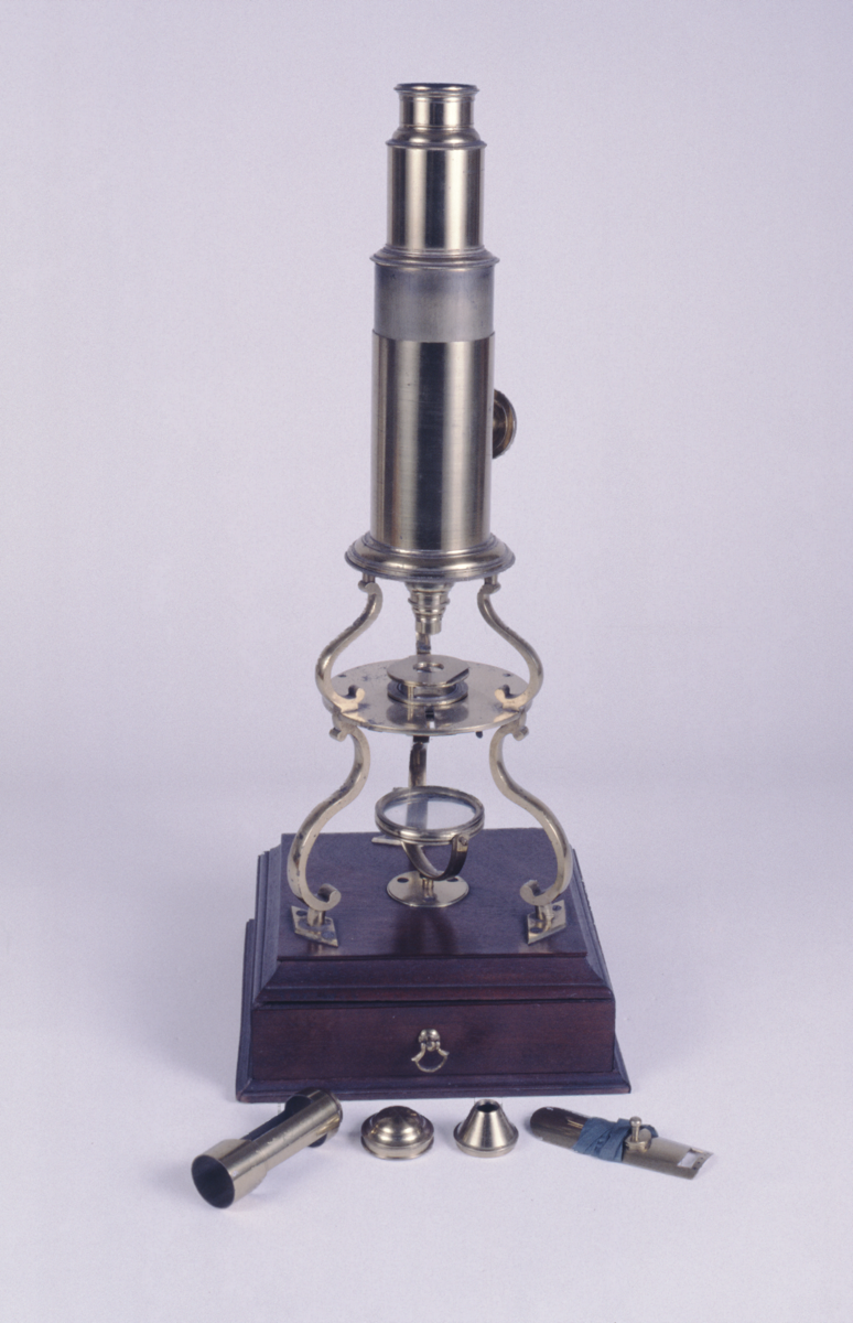
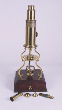
compound microscope, culpeper type, by Silberrad, English, 1801-1833 (c)
Origin
England; London; Aldgate
Maker
Silberrad
Class
microscopes
Earliest Date
1801
Latest Date
1833
Inscription Date
Material
metal (brass); wood (mahogany, other); glass; ivory
Dimensions
height 380mm; depth 159mm; breadth 161mm
Special Collection
Robert Whipple collection
Provenance
Inscription
‘SILBERRAD
Aldgate London’
Description Notes
Brass; square mahogany base with soft wood core; sliding drawer for accessories; 3 scroll legs on diamond feet; circular stage with fittings for frog plate, stage forceps etc.; 3 scroll legs to collar with rack to knurled pinion on body; screw fit field lens mount and screw fit eyepiece. Swinging concave mirror on box lid; 5 objectives marked ‘1-5’; 1 ivory talc and ring box; frog plate; sprung stage; five 4-object ivory slides with long hand list of specimens; tweezers; lieberkuhn in brass box; live object glass tube and wire; cone; speculum (lieberkuhn) holder.
References
Events
Description
The 'culpeper' type microscope
Edmund Culpeper, an instrument maker and engraver of outstanding quality developed the tripod compound microscope in the early 18th century. He mounted the body on two tiers with tripod legs and added a mirror below the stage (the part that holds the specimen). This made it possible to illuminate the specimen from below without having to hold the instrument to the light.
The 'Culpeper' form of microscope quickly became immensely popular and the design was copied by all the leading instrument makers of the 18th century. The materials used gradually changed as the century progressed, from leather, wood and brass, to all brass by 1800.
More on compound microscopes
Culpeper type microscopes are compound microscopes. The compound microscope was developed during the 17th Century and was closely related to the refracting telescope. Its popularity increased after the publication in 1665 of Robert Hooke’s (1635-1703) Micrographia. Micrographia contained detailed pictures, never before seen, of insects magnified using a compound microscope.
A compound microscope uses two or more lenses. The lenses are held at certain distances from each other and are mounted inside a rigid tube. The tube was usually made from pasteboard, ivory, or most commonly, brass. The basic compound microscope magnifies an image in two stages -
Stage one: Light from a mirror is reflected up through the specimen into a powerful objective lens.
Stage two: The image produced by the objective lens is magnified again by the eye lens, which works like a simple magnifying lens.
The first compound microscope consisted of a simple barrel which would have been held up to the light. Later developments ensured that the compound microscope had a stable base, usually a brass stand and a side pillar.
In the 17th Century, the compound microscope had some serious drawbacks which made it easier to use a simple microscope (which have only one lens) instead. The image produced by a compound microscope was often affected by two types of aberration, known as chromatic and spherical. These aberrations caused blurring to the image (spherical) and the edge of the specimen to colour (chromatic). Chromatic aberration was removed at the end of the 18th Century by Harmanus van Deijlan, an instrument maker in Amsterdam. In 1830, spherical aberration was overcome by Joseph Lister who developed the achromatic lens. Achromatic lenses became widely used in microscopes in the 1850s and are still used today.
30/08/2006
Created by: Corrina Bower; updated by Ruth Horry on 30/08/2006
FM:42947
Images (Click to view full size):