Accession No
0937
Brief Description
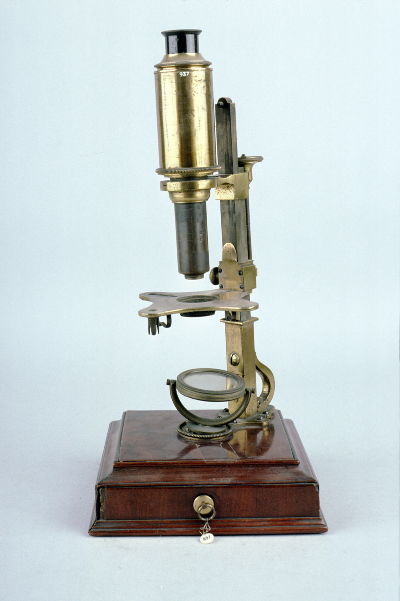
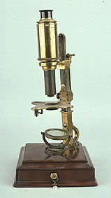
compound microscope, side pillar type, by Scatliff, 1760 (c)
Origin
England; London
Maker
Scatliff
Class
microscopes
Earliest Date
1760
Latest Date
1760
Inscription Date
Material
wood (mahogany, softwood); glass; metal (brass); hide (leather); ivory
Dimensions
height 350mm; depth 170mm; breadth 170mm
Special Collection
Robert Whipple collection
Provenance
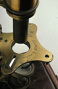
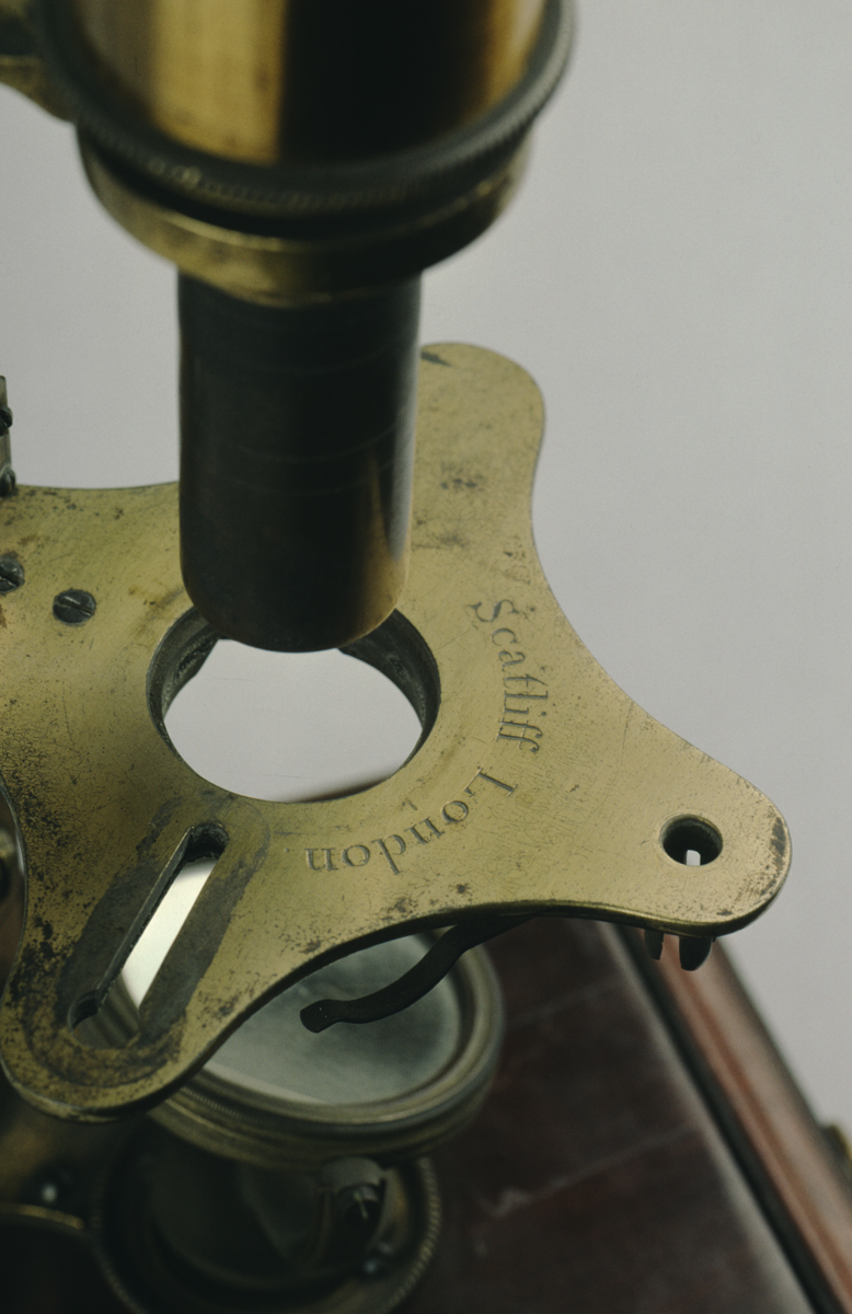
Inscription
‘Scatliff London’
Description Notes
Mahogany and softwood fitted box base swinging concave mirror on brass base [not original]; double rectangular pillar with brace; cruciform stage with slots for stage forceps etc.; coarse focus by sliding shoes on column with clamp; fine focus screw; pillar graduated 1-5; collar to body; push fit; long screw fit snout; single draw tube with field lens and eyepiece in brass cell; eye shade.
Accessories: 6 objectives ‘1-6’, frog plate; lieberkuhn holder; cone; talc and ring box; stage forceps; lieberkuhn; live box; leather case with 4 ivory slides and one brass live slide.
Case (now missing).
References
Events
Description
The compound microscope was developed during the 17th Century and was closely related to the refracting telescope. Its popularity increased after the publication of Robert Hooke’s (1635-1703) Micrographia in 1665. Micrographia contained detailed pictures, never before seen, of insects magnified using a compound microscope.
A compound microscope uses two or more lenses. The lenses are held at certain distances from each other and are mounted inside a rigid tube. The tube was usually made from pasteboard, ivory, or most commonly, brass. The basic compound microscope magnifies an image in two stages -
Stage one: Light from a mirror is reflected up through the specimen into a powerful objective lens.
Stage two: The image produced by the objective lens is magnified again by the eye lens, which works like a simple magnifying lens.
The first compound microscope consisted of a simple barrel which would have been held up to the light. Later developments ensured that the compound microscope had a stable base, usually a brass stand and a side pillar.
In the 17th Century, the compound microscope had some serious drawbacks which made it easier to use a simple microscope (which have only one lens) instead. The image produced by a compound microscope was often affected by two types of aberrations known as chromatic and spherical. These aberrations caused blurring to the image (spherical) and the edge of the specimen to colour (chromatic). Chromatic aberration was removed at the end of the 18th Century by Harmanus van Deijlan, an instrument maker in Amsterdam. In 1830, spherical aberration was overcome by Joseph Lister, who developed the achromatic lens. Achromatic lenses became widely used in microscopes in the 1850s and are still used today.
Created by: Corrina Bower
FM:42952
Images (Click to view full size):