Accession No
1814
Brief Description
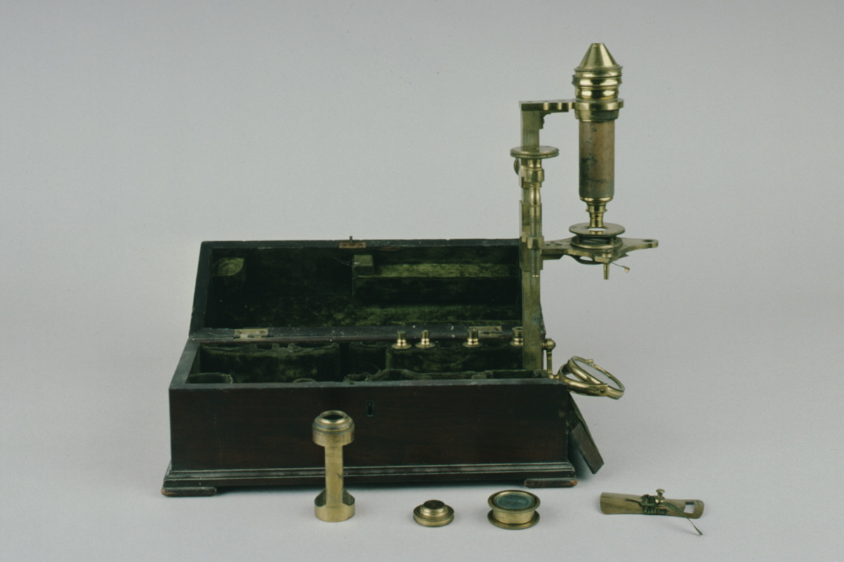
compound microscope, chest type, by Jesse Ramsden, 1790 (c)
Origin
England; London
Maker
Ramsden, Jesse
Class
microscopes
Earliest Date
1790
Latest Date
1790
Inscription Date
Material
wood (mahogany); cloth (velvet); metal (brass); glass
Dimensions
box height 297mm; depth 135mm; breadth 152mm
Special Collection
Heywood collection
Provenance
Purchased from the H.Heywood collection under estate duty exemption benefit with the assistance of a Science Museum grant-in-aid.
Inscription
‘Ramsden London’ (signed round body mount)
Description Notes
compound microscope, chest type, by Jesse Ramsden, c. 1790.
Mahogany box lined with green velvet. Hinged lid and hinged end to allow brass pillar to hinge out of box; swinging concave mirror on articulated arm at base of pillar; cruciform stage, with fittings for stage forceps etc. on shoe round pillar with brass focussing screw and clamp; arm at head for body; push fit snout; (no eye piece collar or eyepiece extant); 5 (out of 6) screw fit objectives; lieberkuhn holder; lieberkuhn in brass case; frog plate; cone; live box; bonnani type stage.
References
Events
Description
The compound microscope was developed during the 17th Century and was closely related to the refracting telescope. Its popularity increased after the publication of Robert Hooke’s (1635-1703) Micrographia in 1665. Micrographia contained detailed pictures, never before seen, of insects magnified using a compound microscope.
A compound microscope uses two or more lenses. The lenses are held at certain distances from each other and are mounted inside a rigid tube. The tube was usually made from pasteboard, ivory, or most commonly, brass. The basic compound microscope magnifies an image in two stages -
Stage one: Light from a mirror is reflected up through the specimen into a powerful objective lens.
Stage two: The image produced by the objective lens is magnified again by the eye lens, which works like a simple magnifying lens.
The first compound microscope consisted of a simple barrel which would have been held up to the light. Later developments ensured that the compound microscope had a stable base, usually a brass stand and a side pillar.
In the 17th Century, the compound microscope had some serious drawbacks which made it easier to use a simple microscope (which have only one lens) instead. The image produced by a compound microscope was often affected by two types of aberrations known as chromatic and spherical. These aberrations caused blurring to the image (spherical) and the edge of the specimen to colour (chromatic). Chromatic aberration was removed at the end of the 18th Century by Harmanus van Deijlan, an instrument maker in Amsterdam. In 1830, spherical aberration was overcome by Joseph Lister, who developed the achromatic lens. Achromatic lenses became widely used in microscopes in the 1850s and are still used today.
FM:42998
Images (Click to view full size):