Accession No
0598
Brief Description
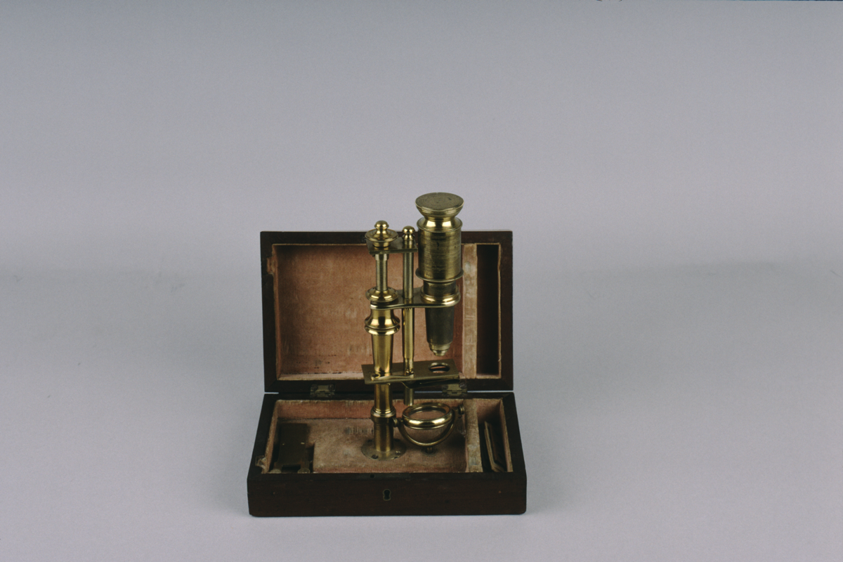
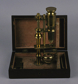
compound microscope, side pillar type; [Dutch]; 2/2 18th century
Origin
[Holland] (Netherlands)
Maker
Class
microscopes
Earliest Date
1750
Latest Date
1800
Inscription Date
Material
wood (mahogany); cloth (velvet); metal (brass); ivory
Dimensions
box length 216mm; breadth 142mm; height 70mm
Special Collection
Robert Whipple collection
Provenance
Purchased from T.H. Court in 03/1930.
Inscription
Description Notes
Mahogany box with marquetry inlay in lid lined with pink velvet; brass plate in base; brass column screws to plate; second rod parallel to first joined by rectangular stage with spring clips below and circular aperture; collar with screw thread and knurled head raises bar holding body; push fit body, tapered snout with field lens in brass cell and screw fit compound eyepiece with screw-fit dust cover rectangular plate at top holds the two columns together, one with screw fit finial; objective marked ‘3’; 2 ivory slides 1 brass live slide casing; spare stage plate [frog plate or unknown function].
References
Events
Description
This type of microscope was first designed by Henry Baker, a microscopist, and John Cuff, an instrument maker, in 1743. By mounting the stage on a side pillar the instrument became easier to use, with the operating parts much more accessible than in previous designs. The focus was controlled by a finely tuned screwthread, and was thus made far more accurate.
More on compound microscopes
The Compound microscope was developed during the 17th Century and was closely related to the refracting telescope. Its popularity increased after the publication in 1665 of Robert Hooke’s (1635-1703) Micrographia. Micrographia contained detailed pictures, never before seen, of insects magnified using a compound microscope.
A compound microscope uses two or more lenses. The lenses are held at certain distances from each other and are mounted inside a rigid tube. The tube was usually made from pasteboard, ivory or most commonly brass. The basic compound microscope magnifies an image in two stages -
Stage One: Light from a mirror is reflected up through the specimen into a powerful objective lens.
Stage Two: The image produced by the objective lens is magnified again by the eye lens, which works like a simple magnifying lens.
The first compound microscope consisted of a simple barrel which would have been held up to the light. Later developments ensured that the compound microscope had a stable base, usually a brass stand and a side pillar.
In the 17th Century the compound microscope had some serious drawbacks which made it easier to use a simple microscope (which have only one lens) instead. The image produced by a compound microscope was often affected by two types of aberrations known as chromatic and spherical. These aberrations caused blurring to the image (spherical) and the edge of the specimen to colour (chromatic). Chromatic aberration was removed at the end of the 18th Century by Harmanus van Deijlan, an instrument maker in Amsterdam. In 1830, spherical aberration was overcome by Joseph Lister who developed the achromatic lens. Achromatic lenses became widely used in microscopes in the 1850s and are still used today.
FM:43013
Images (Click to view full size):