Accession No
6376
Brief Description
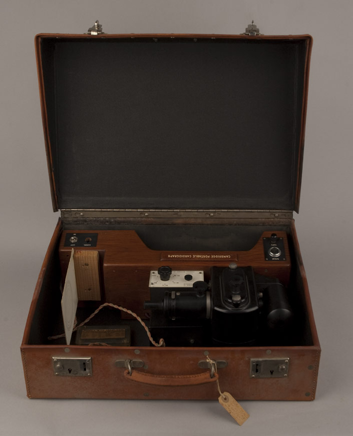
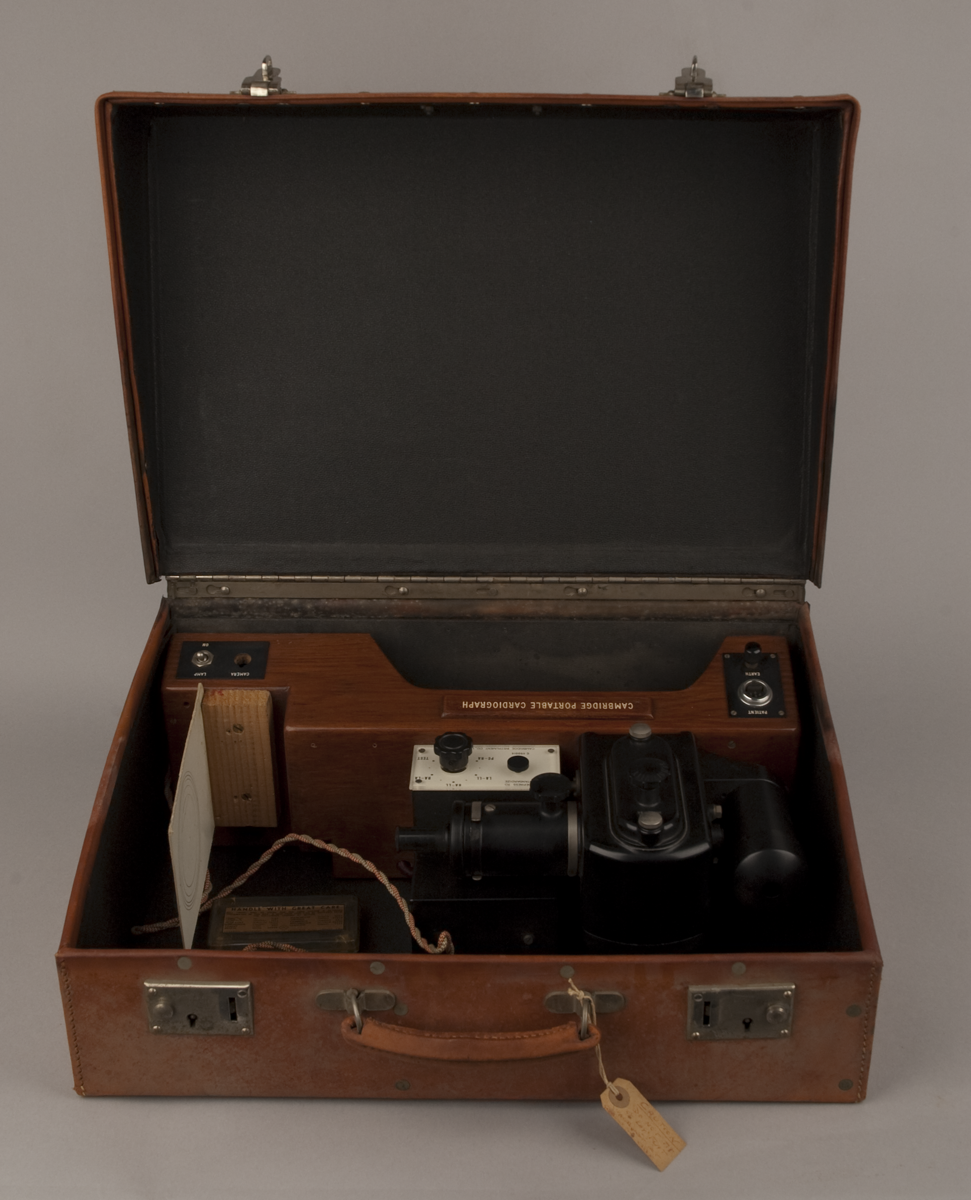
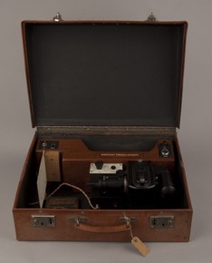
suitcase Portable electrocardiograph (ECG), by Cambridge Instrument Company Ltd., English, 1948
Origin
England; Cambridge
Maker
Cambridge Instrument Company Ltd.
Class
medical; electrical
Earliest Date
1948
Latest Date
1948
Inscription Date
Material
hide (leather); wood (2 types); metal (steel, brass, copper, at least 2 white metals); plastic (perspex, at least 2 other types); cloth (2 types); glass; paper (card); stone
Dimensions
case length 505mm; breadth 210mm; depth 347mm
Special Collection
Provenance
Inscription
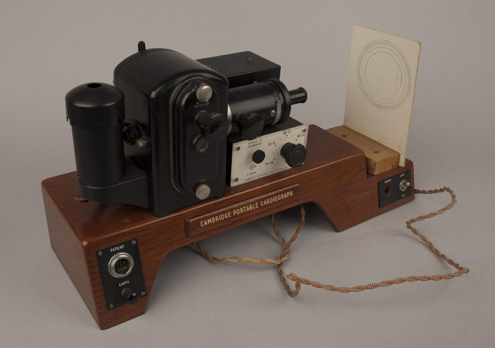
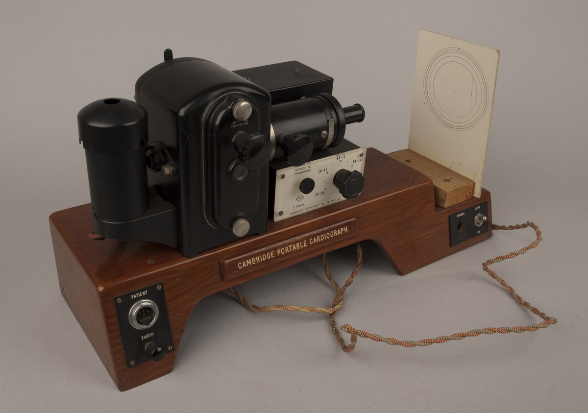
‘CAMBRIDGE INSTRUMENT CO., LTD. ENGLAND’ (on front plate)
‘CAMBRIDGE PORTABLE CARDIOGRAPH’ (on base)
Description Notes
Portable electrocardiograph (ECG), by The Cambridge Instrument Co., England, 1948.
Wooden base supports camera, standardizing switch, lead selector switch, time marker, string galvanometer, and projection lamp. Sensitivity and focussing knobs on front. Main switch which can be set to ‘PC-RA’, ‘LA-LL’, ‘RA-LL’, ‘RA-LA’, ‘TEST’, and ‘OFF’. Lead which would have gone to transformer (missing, would normally have screwed into lid of case). On/off switches for camera and lamp. connection point for electrodes, and earth terminal. Vertical cardboard plate set at one end with concentric circles drawn on it in pencil. The whole fits into a steel framed leather case with reinforced carrying handle. Lid of case held by detachable hinges so that it may be removed. Case contains 2 spare fibre-holders in perspex boxes.
Condition good; incomplete
References
Events
Description
Electrocardiographs (ECGs) are medical instruments used to measure the rhythm at which the heart contracts and the relative strength of different parts of the heart muscle. Electrical impulses in the heart are created at the sinoatrial node – a ‘pacemaker’ tissue in the right atrium – and travel to the heart muscle. On receiving this impulse, a contraction of the muscle fibres is induced and blood is pumped. Electrodes placed on the skin of a patient measure the voltage at different sides of the heart muscle and the ECG subsequently measures the difference in voltage between pairs of electrodes. The product of these measurements is a cardiogram – a record that displays an interpretation of the electrical activity of the heart over time. ECG’s are particularly useful for the diagnosis of irregular heartbeat rhythms caused by damage to the conductive tissue in the heart or by an inadequacy or abundance of electrolytes, such as Potassium and other dissolved salts. However, it is not capable of measuring all aspects of muscle activity in the heart and is ineffective in measuring pumping ability in comparison to echocardiographic methods.
Although technologies that recorded electrical traces of the heartbeat had been developed at the end of the nineteenth-century, Willem Eithoven’s String Galvanometer was found to be capable of more sensitive measurements than its predecessors. Eithoven characterised several ECG measurements, described electrocardiographic features of several cardiovascular disorders, and was later awarded the Nobel Prize in Medicine for his discovery.
The Cambridge Scientific Instrument Company developed their first ECG in 1912 based on Eithoven’s design. However, the instrument covered a floor space of 6ft by 3ft and weighed 336lb. It was far from portable and one of the most astonishing elements of the history of the ECG is the shift from a cumbersome piece of equipment used by the hospital specialist to the portable instrument of the general clinician. This particular ECG was produced in England in 1948.
18/09/2009
Created by: M.A. Coxhead on 18/09/2009
FM:43916
Images (Click to view full size):