Accession No
1426
Brief Description
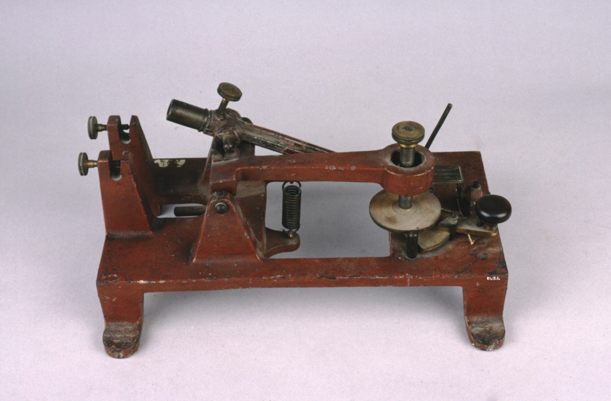
microtome, rocking, by Cambridge Scientific Instrument Company Ltd., English, circa 1895
Origin
England; Cambridge
Maker
Cambridge Scientific Instrument Company Ltd.
Class
laboratory apparatus; biology; microscopes
Earliest Date
1895
Latest Date
1895
Inscription Date
Material
metal (iron, brass, steel)
Dimensions
length 365mm; breadth 245mm; height 160mm
Special Collection
Provenance
Transferred from the Pathology Laboratory, University of Cambridge, in 1963.
Inscription
‘CAMBRIDGE SCIENTIFIC
INSTRUMENT COMPANY
LIMITED’ (on brass plaque)
Description Notes
Microtome, rocking; made by the Cambridge Scientific Instrument Company; circa 1895.
Cast iron base with splayed feet finished in red paint; cast verticals with brass knurled screw clamps for knife (missing); cast uprights for pivotted support for rocking arm; rocking arm with steel axis and brass block with screw clamp (no steel shaft into the arm as on Wh: 1363), support on steel screw with brass cogged wheel operated by a handle on brass shaft with spring steel cog turning the brass wheel, and connected by pulley to the rocking arm.
References
Events
Description
A microtome is a laboratory instrument used to cut extremely thin slices of material, called sections. These are usually cut from specimens of human or animal tissue (embedded in a soft material like paraffin wax), and are produced for inspection under a microscope. The “rocking” type of microtome was designed by Charles Darwin’s son, Horace Darwin. Horace co-founded the Cambridge Scientific Instrument Company with Albert George Dew-Smith in 1881, and the firm began manufacturing Horace’s microtome design from 1885. This design, with updates, continued to be produced well into the second half of the twentieth century. As a Cambridge Instruments sales catalogue boasted, “simplicity of operation makes it an ideal instrument for the use of students or for routine work and it has become the standard microtome for general use in laboratories all over the world.”
This particular model could cut sections between 0.002mm and 0.024mm thick.
14/03/2014
Created by: Joshua Nall on 14/03/2014
FM:44093
Images (Click to view full size):