Accession No
4094
Brief Description
portable electrocardiograph (ECG), by Cambridge Instrument Company Ltd., English, 1935
Origin
England; Cambridge
Maker
Cambridge Instrument Company Ltd.
Class
medical
Earliest Date
1935
Latest Date
1935
Inscription Date
Material
metal (brass, iron, at least 4 others); plastic (ebonite, bakelite, perspex, at least 2 others); cloth; glass; rubber
Dimensions
length 615mm; breadth 293mm; height 312mm
Special Collection
Cambridge Instrument Company Collection
Provenance
Donated by the Cambridge Instrument Company.
Inscription
‘Cambridge Instrument Co. Ltd., England’ (on camera)
‘CAMBRIDGE PORTABLE
ELECTROCARDIOGRAPH
[CIC logo]
THE PROPERTY OF
DR S. S. SUZMAN,
84 HARLEY ST.
LONDON W.1’ (on case)
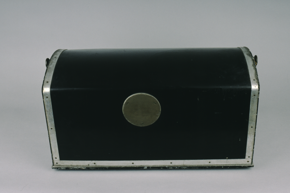
Description Notes
portable electrocardiograph (ECG), by the Cambridge Instrument Company, 1935.
Black-painted iron base. Camera mounted at rear with horizontallly mounted film canister, brass shutter release. The shutter itself is in 3 sections, each moved by metal knob. Scale above shutter divided 40 - 0 - 40 numbered by 10 subdivided to 1. Front section of instrument carries dial for galvanometer divided 0 - 1 Volts numbered by 0.5. Front also carries adjustment knob for rheostat, switch which moves between ‘I.MV.’, ‘0’, ‘SHUNT’ and ‘SHORT’. One further knob of unknown purpose. Central section carries black-painted casing around lamp, galvanometer with sensitivity adjustment knob and focussing optics. The display is projected onto a mirror angled to bounce the image onto a mirror at the front of the instrument, which directs the light onto the shutter. Central section also carries stand for spare film canisters, under which is a compartment for storing canisters and electrodes. Black plastic case bound with white metal, with telescopic handles.
References
Events
Description
Electrocardiographs are medical instruments that measure the rhythm of heart contractions and the relative strength of different parts of the heart muscle. This portable electrocardiograph had been the property of Dr S. S. Suzman who worked in the Cardiographic Department of Guy’s Hospital during the 1940s. With a team of researchers, he studied and published on: cyanotic congenital heart disease in children; pericardial effusion, or an accumulation of fluid in the sac membrane that contains the heart; the narrowing of the aorta, or the left ventricle that distributes oxygenated blood to the body. The death of Dr S. S. Suzman was noted at the Forty-third Annual General Meeting of the British Cardiac Society held in Liverpool, 8–9 April 1965.
06/11/2013
Created by: Allison Ksiazkiewicz on 06/11/2013
FM:44337
Images (Click to view full size):