Accession No
5240
Brief Description
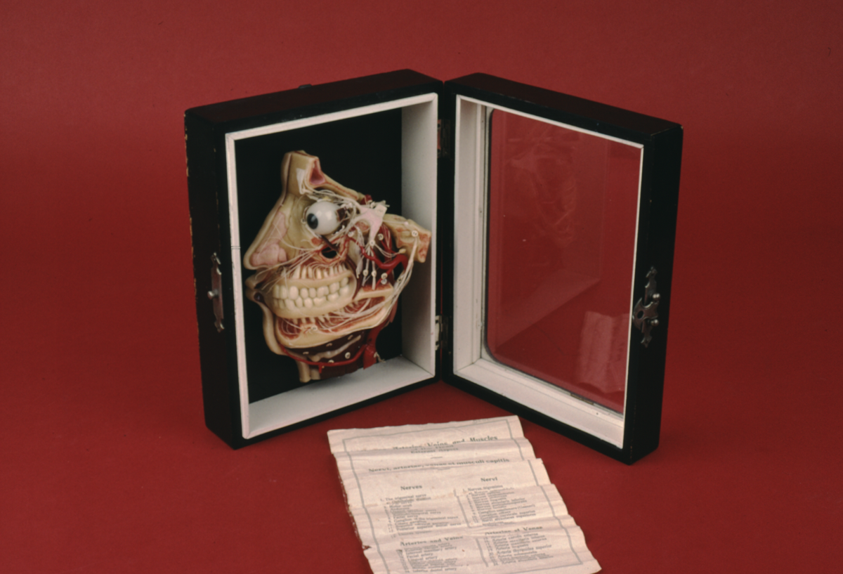
Wax anatomical model of the human head, by Lehrmittelwerke, 2/2 19th C
Origin
Berlinische Verlagsanstalt G. m. b. H; Berlin N. W. 23; Germany
Maker
Lehrmittelwerke
Class
physiology
Earliest Date
1850
Latest Date
1900
Inscription Date
Material
wood; glass; metal (white metal); wax; thread; paper
Dimensions
width 192 mm; height 252 mm; breadth 78 mm
Special Collection
Provenance
Purchased from David Burns, Unit 12, 284 Westbourne Grove, London, W11, England.
Inscription
‘Lehrmittelwerke
Berlinishce Verlagsanstalt
G.m.b.H.
BERLIN N.W. 23’ (paper label on back of case)
Description Notes
Wax profile model of the human head, without the upper part of the skull. Shows major blood vessels, eye, teeth with nerves, and sinus cavities. Various parts are numbered from 1 to 39. Mounted in glazed wooden display case with white metal hinges and clip fastener.
Accompanying paper key gives names of numbered items, in sections ‘Nerves’, ‘Arteries and Veins’, ‘Muscles’ and ‘Bones, cavities, etc.’. Information is provided in both English and Latin.
A paper label on the back of the box warns that the model must be kept out of direct sunlight.
Condition good; complete.
References
Events
Description
This wax models shows a profile of a human head without the upper part of the skull. The major blood vessels, eye, teeth and nerves, as well as sinus cavities are exposed. In the nineteenth century, the importance of performing dissection during medical training increased, and by 1875 it had become mandatory for British medical students to undertake dissection. Confronted with a shortage of human remains for performing dissections, nineteenth-century model-makers began to produce life-like examples of anatomy that supported medical teaching. Anatomical models facilitated a physician’s training, as they were important objects for familiarizing and visualizing internal human anatomy.
18/02/2014
Created by: Allison Ksiazkiewicz on 18/02/2014
FM:45386
Images (Click to view full size):