Accession No
5805
Brief Description
anatomical model of a human ear in papier-mâché and plaster, late 19th to early 20th Century
Origin
Maker
unknown
Class
physiology; sound
Earliest Date
1850
Latest Date
1950
Inscription Date
Material
wood; plaster; paper (papier-mâché); metal
Dimensions
height 245mm; width 350mm; depth 180mm
Special Collection
Provenance
Purchased on or before 31/10/2000.
Inscription
Description Notes
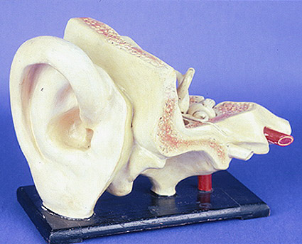
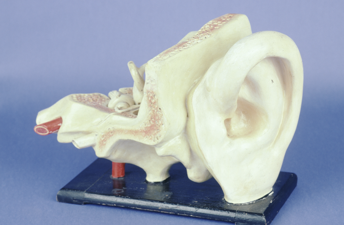
anatomical model of a human ear in papier-mâché and plaster on a wooden base. Late 19th / early 20th century. No maker's mark.
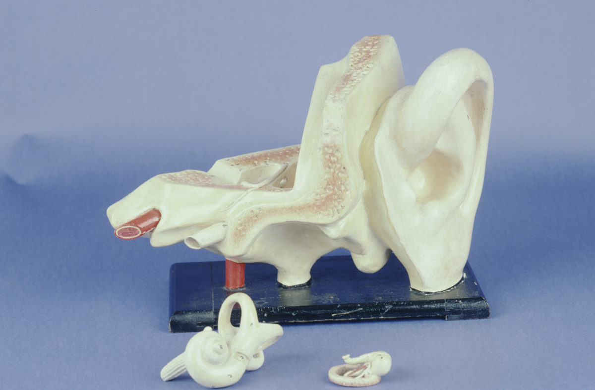
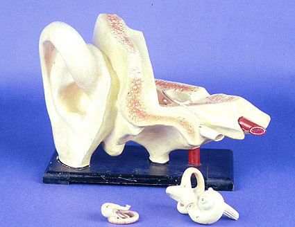
Two pieces can be removed, the ear drum and the cochlea. The parts of the ear are labelled with numbers and it is likely a key would have been provided.
References
Events
Description
This papier-mâché model represents a human ear. It comes apart to reveal the ear drum and other inner structures. Models like this played an important part in medical training.
Ears have a shell-like shape to collect sound waves and funnel them down to the ear drum. The vibrations of the very thin membrane of the ear drum create signals for your brain to interpret.
People experience sound in different ways. Some people are very sensitive to noise, while others need the stimulation of lots of sound. Young people can hear higher-pitched sounds than older people. Some people may not hear sound with their ears, but can feel the vibrations of music and rhythms through their bodies.
22/07/2024
Created by: Hannah Price on 22/07/2024
Description
This papier-mâché model of the human ear has two pieces that can be removed to better demonstrate anatomical arrangement: the tympanic membrane, or ear-drum, and the cochlea. In the early nineteenth century, model makers began to experiment with different materials to produce durable and inexpensive models of human anatomy. Traditional wax models were too fragile and expensive to be handled for demonstration and could not be disassembled. Throughout this period, the importance of performing dissection during medical training increased. By 1875 it had become mandatory for British medical students to undertake dissection. Confronted with a shortage of human remains, anatomical modals facilitated a physician’s training and became important objects for familiarizing and visualizing internal human anatomy.
18/02/2014
Created by: Allison Ksiazkiewicz on 18/02/2014
FM:46210
Images (Click to view full size):