Accession No
6185
Brief Description
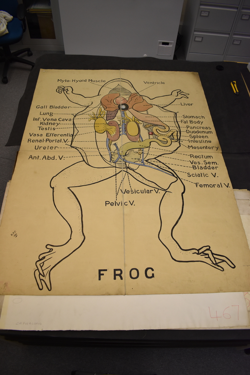
zoological wall chart depicting the frog, attributed to K. P. P., English, 1911
Origin
England; Cambridge; Department of Zoology
Maker
K. P. P. [attributed]
Class
natural history; physiology; prints
Earliest Date
1911
Latest Date
1911
Inscription Date
1911
Material
paper; cloth (canvas)
Dimensions
1173mm; 1595mm
Special Collection
Provenance
Transferred by an individual at the Department of Zoology, University of Cambridge, on or before 05/06/2006.
Inscription
FROG
K.P.P. 1911
Description Notes
Zoological wall chart depicting the ventral anatomy of a frog. The frog appears to be drawn from an actual dissection. Organs labelled include the ventricle, liver, stomach, fat body, pancreas, duodenum, spleen, intestine, mesentery, rectum, ves. sem., bladder, sciatic vein, femoral vein, vesicular vein, pelvic vein, anterior abdominal vein, ureter, renal portal vein, vasa efferentia, testis, kidney, inferior vena cava, lung, gall bladder and mylo-hyoid muscle.
The image is hand painted and in colour.
The initials K.P.P. appear in the left hand bottom corner along with the date 1911.
References
Events
Description
This diagrams is part of a collection of around 100 zoological illustrations which were donated from the Department of Zoology in 2006. The diagrams were used for teaching, and were hand drawn. All of the diagrams are very large, so that in lectures all the students could easily see them.
[Label written by Rosanna Evans, year 10 work experience student]
Created by: Rosanna Evans, year 10 work experience student
FM:46653
Images (Click to view full size):