Accession No
6194
Brief Description
papier mâché and glass anatomical model of a human eye, by Louis Thomas Jérôme Auzoux, French, 1900 (c)
Origin
France; Paris
Maker
Auzoux, Louis Thomas Jérôme
Class
demonstration; physiology; biology; medical
Earliest Date
1850
Latest Date
1950
Inscription Date
Material
paper (papier mâche); glass
Dimensions
length 345mm; width 285mm; height 155mm
Special Collection
Provenance
Purchased from Marc-Andre & Marlyse Perret, Antiquites Scientifiques, 19 Rue du Perron, 1211, Geneve, Switzerland on or before 15/11/2007.
Inscription
Dr AUZOUX S.A.
PARIS FRANCE
Description Notes
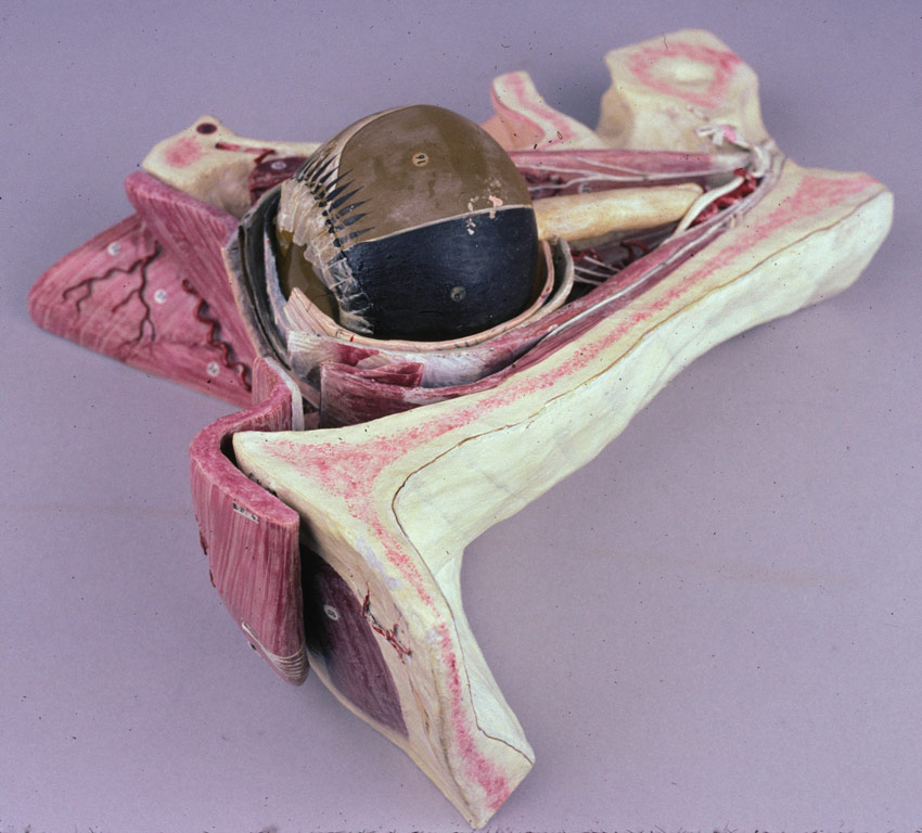
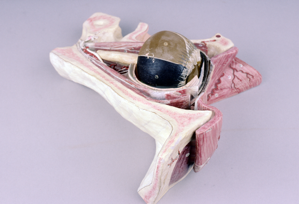
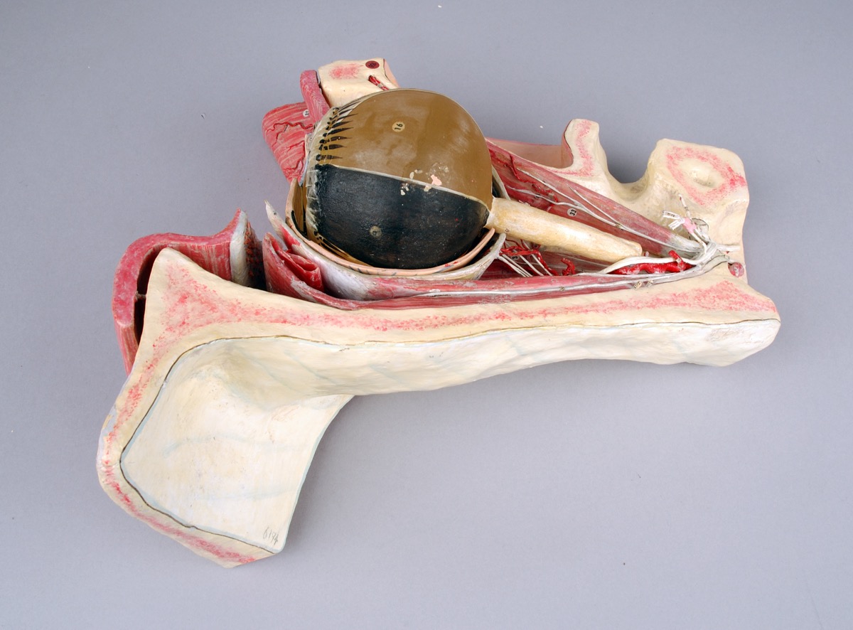
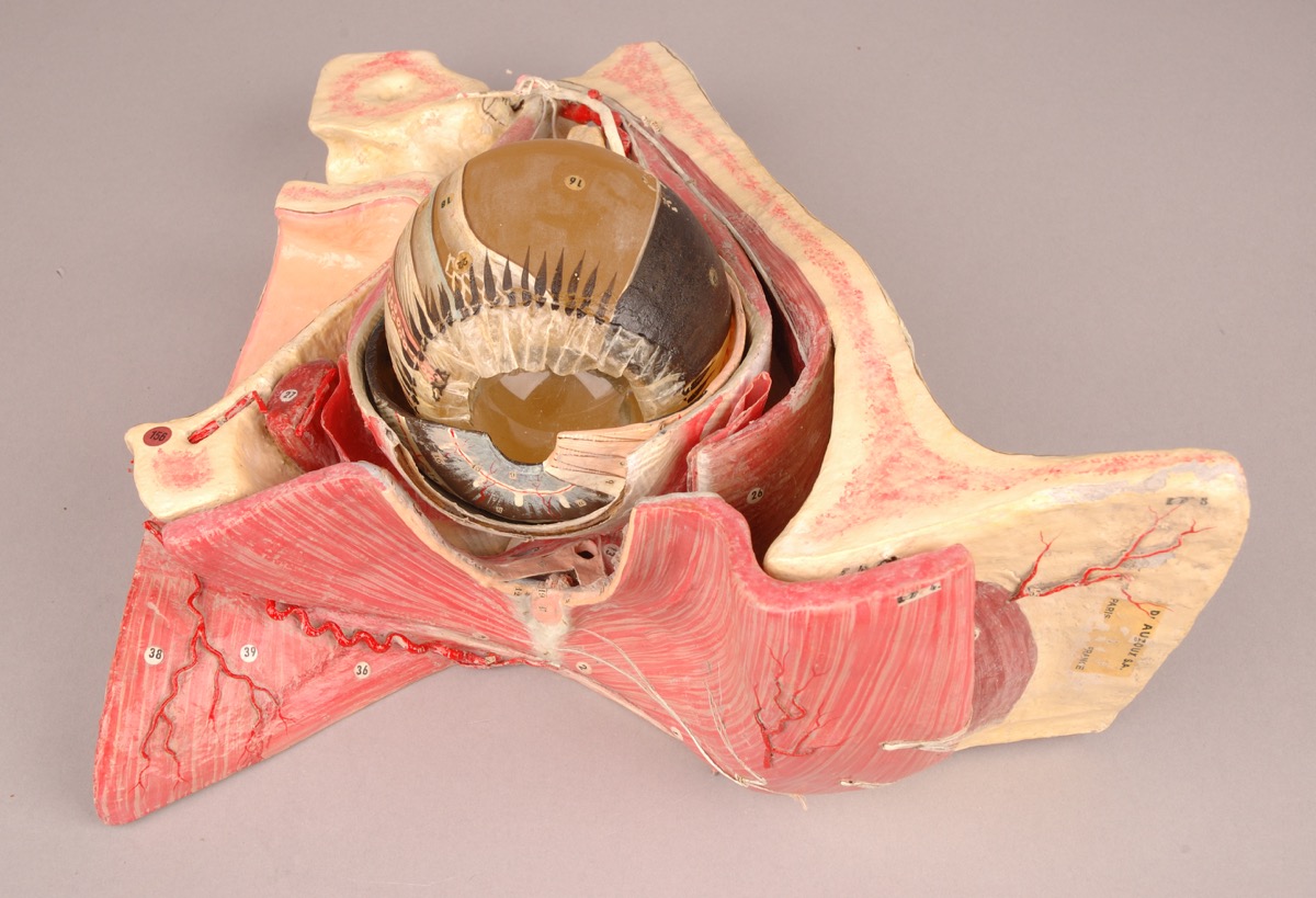
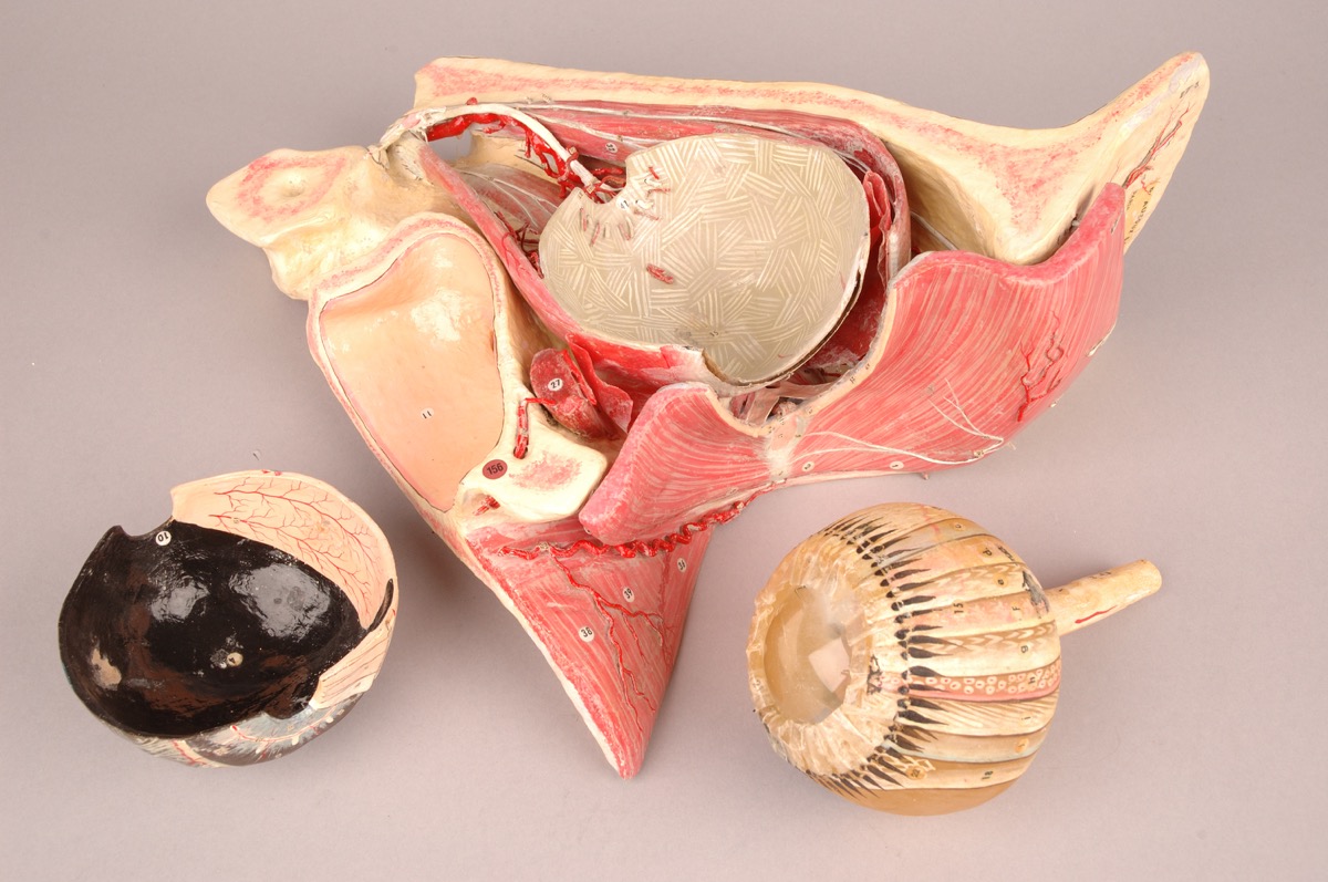
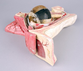
Anatomical model of human eye, in papier mache and glass, by “Auzoux S.A. Paris”, French, c. 1900.
Anatomical model showing ‘cut-away’ side view of eye socket and surrounding facial musculoskeletal features, which sits on its side, for top-down viewing. The base consists of section of skull plus muscle structure in which the eyeball sits. This structure includes numerous features (labelled with numbers) including veins, arteries and other muscle or nerve strands.
The eyeball itself consists of painted glass (also with labels) plus papier mache optic nerve at rear. The cornea is represented by a loose glass lense. The eyeball structure can be removed entirely from the socket in which it sits and is half encircled by a papier mache outer layer, which can also be removed.
Condition: good/fair (some paint flaking and loss)
References
Events
Description
As the eye was difficult to study in cadavers, it was a particularly popular anatomy to model in the eighteenth and nineteenth centuries. While anatomical models did not completely displace the value of observation through dissection, they were important objects on which surgical techniques could be practiced. The development of new methods for treating cataracts and eye infections such as trachoma, opthalmia neonatorum and syphilis facilitated medical specialization on the eye during the eighteenth and early nineteenth centuries. This model was produced by the French model-maker Louis Thomas Jérôme Auzoux (1797–1880) and represents the eye cut vertically in half and enlarged ten times. The Auzoux eye-model was first made commercially available in 1826; however, the item in the Whipple Museum is most likely an older issue as the labels are printed rather than hand written. Like many of Auzoux’s models, the eye is clastic. All parts can be disassembled as a mode of artificial dissection, making the model a useful tool for medical students and practitioners. For instance, one side of tendon is removable to facilitate clearer viewing of the nerves and blood vessels. The vitreous humour is likewise removable enabling the viewer to study the inner anatomy of the eye.
05/11/2013
Created by: Allison Ksiazkiewicz on 05/11/2013
FM:46662
Images (Click to view full size):