Accession No
6247
Brief Description
Stereoscopic slides of human anatomy, from the Edinburgh Stereoscopic Atlas of Anatomy, by T.C. & E.C. Jack, Scottish, c. 1900
Origin
Edinburgh
Maker
T. C. & E. C. Jack
Class
medical; physiology; prints
Earliest Date
1900
Latest Date
1910
Inscription Date
Material
Paper (cardboard, photographic and one other); Textile (unspecified cloth)
Dimensions
Length 240mm; Width 197mm; Depth 80mm
Special Collection
Provenance
Purchased from Trevor Philip & Sons, 75A Jermyn St., St. James, London, SW1Y 6NP.
Inscription
[6247.1] (left side) THE EDINBURGH STEREOSCOPIC ATLAS OF ANATOMY
SECTION III
CONTENTS - 51 Plates, viz. :-
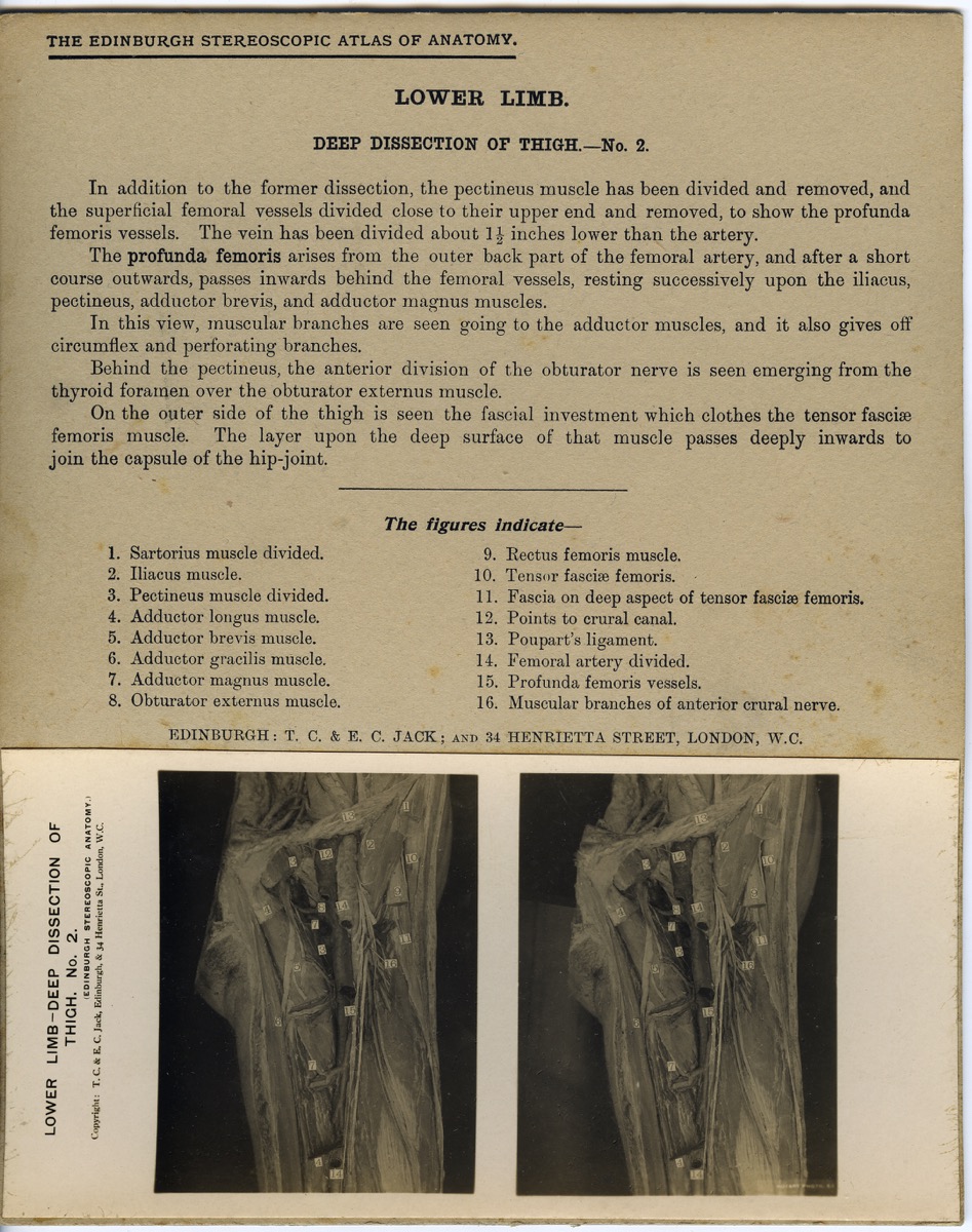
Abdomen and Pelvis.
Lower Limb.
[6247.2] (left side) EDINBURGH STEREOSCOPIC ATLAS OF ANATOMY. SECTION II
CONTENTS - 50 Plates, viz.:-
Abdominal Wall, Nos. 1 to 4.
Inguinal Region, Nos. 1 to 6.
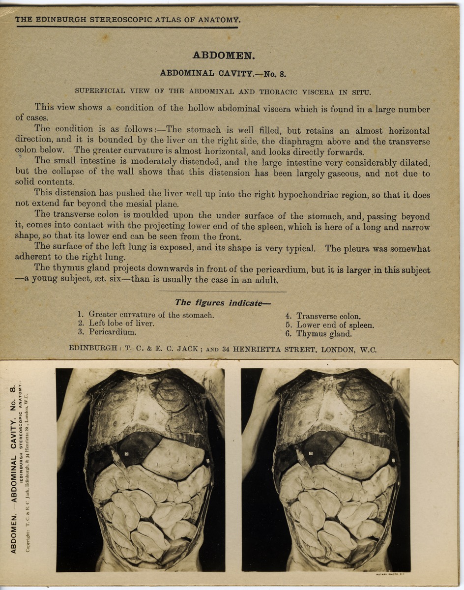
Abdominal Cavity, Nos. 1 to 7.
Viscera, Nos. 1 to 21.
Abdominal Aorta, No. 1.
Pelvis, Nos. 1 to 8.
Bladder, Nos. 1 to 3
(left side) CONTENTS OF THIS SECTION, ABDOMEN
Abdominal Wall, Inguinal Region, Abdominal Cavity, Viscera, Abdominal Aorta, Pelvis, Bladder
[6247.3] (right side) LIST OF VIEW FOR SECTION (subheadings) ABDOMEN AND PELVIS, PELVIS and LOWER LIMB
[6247.4] (right side) EDINBURGH STEREOSCOPIC ATLAS OF ANATOMY
SECTION I
CONTENTS - 50 Plates, viz. :-
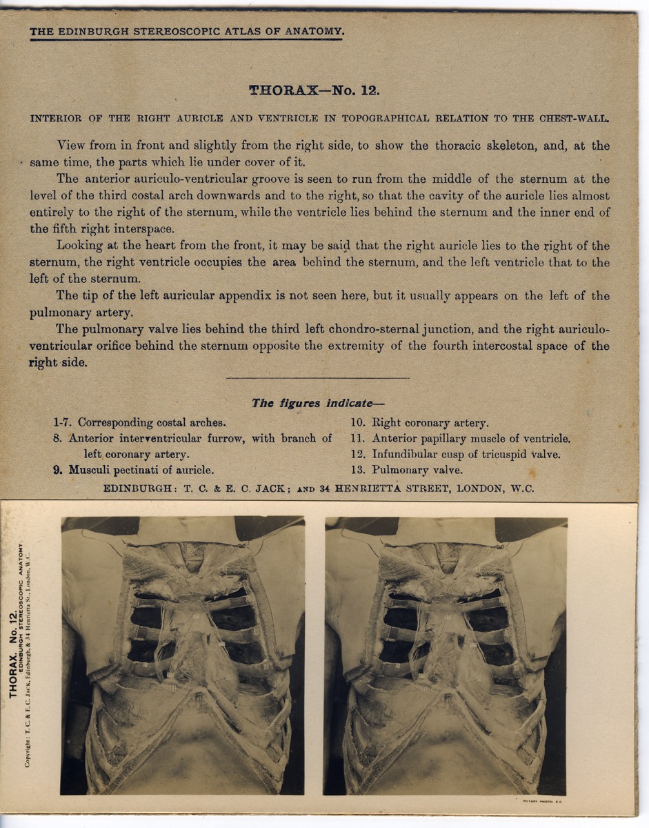
Thorax Nos. 1 to 15
Thorax, Lungs, Nos. 1 to 3
Thorax, Heart and Pericardium, Nos. 1 to 12
Thorax, Mediastina, Nos. 1 to 6
Central. Nervous System - Brain, Nos. 1 to 14
(left side) (List of contents) THORAX and CENTRAL NERVOUS SYSTEM
Description Notes
Four boxes of stereoscopic slides of human anatomy, from the Edinburgh Stereoscopic Atlas of Anatomy, by T.C. & E.C. Jack, Scottish, c. 1900
6247.1
Box is made of medium thickness card covered in blue outer binding. Box is very damaged with right side missing and binding is separating from card beneath.
A length of cloth is attached to the bottom of box to allow the slides to be removed easily.
The left side of the box bears a paper label reading: THE EDINBURGH STEREOSCOPIC ATLAS OF ANATOMY and brief description of contents (see inscription above).
The box contains 53 slides of the abdomen, the pelvis and lower limb. (Note that the paper label on the side of the box states 51 plates enclosed).
Each card slide gives a title and detailed description, figure key and figured stereoscopic photograph (above each photo is written EDINBURGH: T. C. & E. C. JACK: and 34 HENRIETTA STREET, LONDON, W.C.)
6247.2
Box is made of medium thickness card covered in blue outer binding. Box is very damaged with severe rips and tears to the interior and exterior. The left side of the box bears a paper label reading THE EDINBURGH STEREOSCOPIC ATLAS OF ANATOMY and brief description of contents (see inscription above)
The Box contains 53 slides (Note that the inscription states that it only contains 50 slides). The slides show the Abdominal Wall, Inguinal Region, Abdominal Cavity, Viscera, Abdominal Aorta, Pelvis, Bladder
6247.3
Box is made of medium thickness card covered in blue outer binding. Box is very damaged with severe rips and tears to the interior and exterior. All corners of the lid have been mended with acid free tape. Lid has been replaced with what appears to be the cover of a hard back book. It has rough drawings and writing on the inside not relating to the contents of the box. In comparison the bottom half of the box is in much better condition with all four corners intact.
The box contains 50 slides. The slides show the Abdomen and Pelvis, Pelvis and Lower Limb
6247.4
Box is made of medium thickness card covered in blue outer binding. Box is in fair condition, still damaged but much less so than the others. There is a significant bend to the lid which causes difficulty when closing the box. One corner on both lid and and bottom mended with acid free tape. Paper labels on both sides of lid (see inscription above)
The box contains 48 slides. The slides show the Thorax and Central Nervous System.
References
Events
Description
First published in 1905, The Edinburgh Stereoscopic Atlas of Anatomy is the earliest commercial application of stereoscopy to anatomical illustration. It consists of approximately 250 stereoscopy photographs of dissected cadavers, arranged to mimic the process of dissection. Each anatomical view is accompanied by a brief description. The Stereoscopic Atlas of Anatomy grouped the body by regions or topography rather than systematic approaches in popular anatomy textbooks such as Gray’s Anatomy. At the time of publication, a topographical focus in anatomy atlases was increasingly popular, as a way of defending anatomy against the encroachment of experimental physiology and related sciences. In addition to the use of photography, other technological advances such as the cadaveric preservation formalin shaped new approaches to studying and representing the human body. First discovered in 1893, formalin was met with enthusiasm by Edinburgh anatomists who hoped the chemical would ‘effect a total revolution in our knowledge of the abdominal and thoracic viscera.’ Formalin both preserved and hardened cadavers, giving them a rigidity that maintained spatial relationships between tissues throughout dissection. This new chemical technique facilitated in the production of the Atlas’ stereoscopic views.
A stereoscopic photograph is composed of two photographs mounted side by side on a card. The photographs are of the same view, but taken from slightly different angles. When viewed through the stereoscope, the eye only ‘sees’ the image directly in front and the brain combines the two monocular images to produce the illusion of depth and three-dimensionality. In an era of lecture-centered anatomy courses and chronic cadaver shortages, physicians embraced the Atlas when it was introduced. The Atlas was relatively expensive—it cost fifty American dollars for a complete set of stereographs—but it was nonetheless widely circulated within American medical institutions throughout the early twentieth century.
08/07/2014
Created by: Allison Ksiazkiewicz on 08/07/2014
FM:46726
Images (Click to view full size):