Accession No
6598
Brief Description
papier mâché didactic model of a human hand, by Louis Thomas Jérôme Auzoux, French, 1876
Origin
France; Saint-Aubin-d'Écrosville [attributed]
Maker
Auzoux, Louis Thomas Jérôme
Class
physiology; medical; demonstration
Earliest Date
1876
Latest Date
1876
Inscription Date
1876
Material
paper (papier mâché); paint; metal (brass)
Dimensions
51cm long x 30cm wide x 10.5cm tall
Special Collection
Provenance
Purchased from an individual via www.fleaglass.com on or before 27/05/2015.
Inscription
Auzoux [?unreadable?]
fecit anno 1876
Description Notes
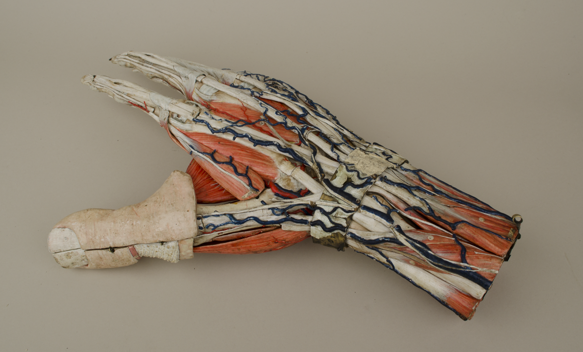
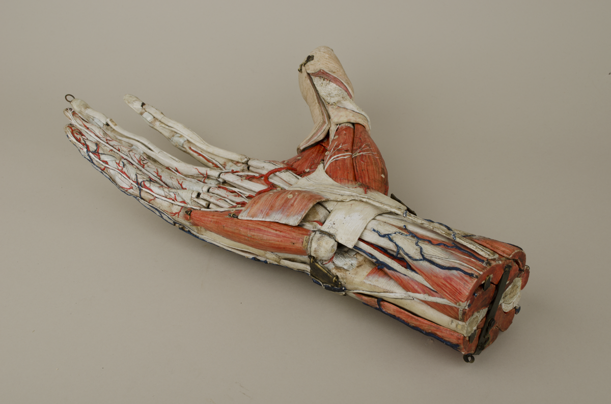
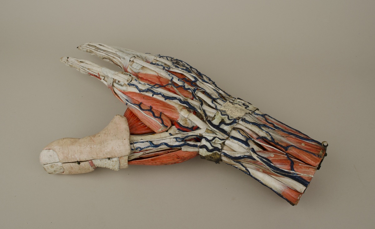
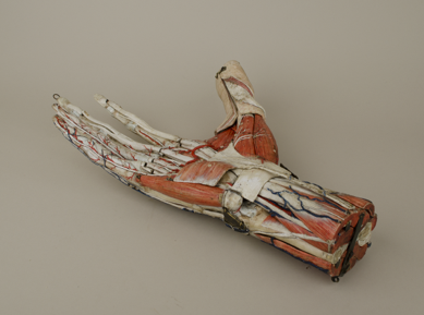
Papier-mâché didactic model of a human hand, by Auzoux, French, 1876.
Papier-mâché model, roughly twice life size, showing the complex anatomy of the human hand, including muscles, nerves, tendons, blood vessels, bones and layers of skin. Composed of around 20 individual removable parts, held together by brass rods, loops, and hooks. Hand-painted in vivid but life-like colours. Covered with hundreds of small circular paper labels carrying numbers.
Thumb of model is encased in a removable hinged section showing flayed layers of skin and subcutaneous fat.
Manuscript inscription on end of wrist “Auzoux [?unreadable?]
fecit anno 1876”.
References
Events
Description
In contrast to an older generation of wax models of fauna anatomy, those produced by Louis Thomas Jérôme Auzoux (1797–1880) enabled a systematic disassembly and reconstruction of the modelled object. Using a new, secret recipe for a tough, durable type of papier-mache, Auzoux produced a wide variety of anatomical, zoological, and botanical didactic models. Intended for university training, the enhanced colouring of the Auzoux models facilitated the identification of anatomy, and an elaborated labelling system accompanied each model. Depending on the example, the identifiable and removable parts could be extensive. For example, the ‘Complete Clastic Human’ model produced by Auzoux in 1858 contained 92 removable pieces on which there were over 2000 labeled details. To enhance the ‘dissection’ experience, a spatula was used to unhinge the clastic pieces of the model in a fashion similar to the surgeon’s scalpel on the dissection table.
This particular model enabled the medical student to carefully study the complex anatomy of the human hand, enlarged to about twice natural size to facilitate close study as the model is taken apart and rebuilt. An Auzoux sales catalogue from the late 19th century explains that the model shows “muscles, tendons and tendinous sheaths disposed so as to show the action of the interosseus and lumbrical muscles indicated by Dr. Duchenne, of Boulogne; the arteries, veins, nerves, corpuscles of Pacini, and a portion of the skin with its various layers.”
05/11/2013
Created by: Allison Ksiazkiewicz on 05/11/2013
FM:47116
Images (Click to view full size):