Accession No
1518
Brief Description
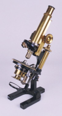
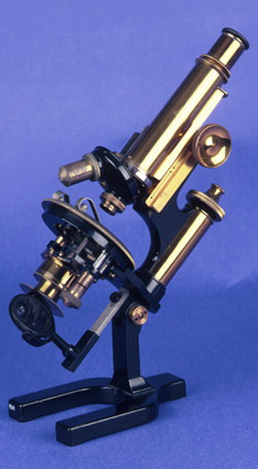
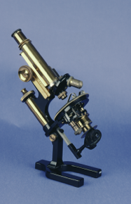
achromatic compound microscope, petrological, by W. Watson and Sons, English, circa 1920
Origin
England; London; 313 High Holborn
Maker
W. Watson and Sons Ltd. J. Swift and Son [objectives]
Class
microscopes
Earliest Date
1920
Latest Date
1920
Inscription Date
Material
metal (brass, oxidised brass, white metal); glass
Dimensions
height 318mm; breadth 100mm; depth 190mm
Special Collection
Provenance
Transferred from the Sedgwick Museum of Earth Sciences, University of Cambridge, in 18/01/1971.
Inscription
‘W. Watson & Sons, Ltd.
313 High Holborn
London.
26727’ (on base)
‘“PRAXIS
PETRO”’ (on base)
‘P-A.
1/4 IN
N.A.O.88
J. SWIFT & SON
LONDON’ (on one objective)
‘J.SWIFT & SON 55619 1 IN N.A.O.16’ (on other objective)
‘SEDG. MUS.
CAMB.
37’ (on body)
‘WOODN. MUS
MO. 14’
Description Notes
Black finish. Brass body and fittings. Horseshoe base. Pivoting limb. Circular stage divided [0] - 360˚, numbered by 10˚, graduated to 1˚, with index. 2 spring clips. Condenser on arm to swing out of optic axis. Polariser on arm also to swing out of axis with divided flange [0] - 360˚, numbered by 30˚, graduated to 10˚. Swinging plano-concave mirror. Cranked limb. Coarse focus screws either side of limb; lever fine focus at head of column with conical cap. Body with draw tube graduated in cm. Push-fit eyepiece. Analyser in box fitting in slot in the nose. Slot for quartz plate. Double nose piece. Two objectives.
Condition: fair; complete
References
Events
Description
The compound microscope developed during the 17th Century and was closely related to the refracting telescope. Its popularity increased after the publication of Robert Hooke’s (1635-1703) Micrographia in 1665. Micrographia contained detailed pictures, never before seen, of insects magnified using a compound microscope.
A compound microscope uses two or more lenses. The lenses are held at certain distances from each other and are mounted inside a rigid tube. The tube was usually made from pasteboard, ivory or most commonly brass. The basic compound microscope magnifies an image in two stages -
Stage one: Light from a mirror is reflected up through the specimen into a powerful objective lens.
Stage two: The image produced by the objective lens is magnified again by the eye lens, which works like a simple magnifying lens.
The first compound microscope consisted of a simple barrel which would have been held up to the light. Later developments ensured that the compound microscope had a stable base, usually a brass stand and a side pillar.
In the 17th Century, the compound microscope had some serious drawbacks which made it easier to use a simple microscope (which have only one lens) instead. The image produced by a compound microscope was often affected by two types of aberrations known as chromatic and spherical. These aberrations caused blurring to the image (spherical) and the edge of the specimen to colour (chromatic). Chromatic aberration was removed at the end of the 18th Century by Harmanus van Deijlan, an instrument maker in Amsterdam. In 1830, spherical aberration was overcome by Joseph Lister, who developed the achromatic lens. Achromatic lenses became widely used in microscopes in the 1850s and are still used today.
FM:40061
Images (Click to view full size):