Accession No
0297
Brief Description
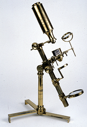
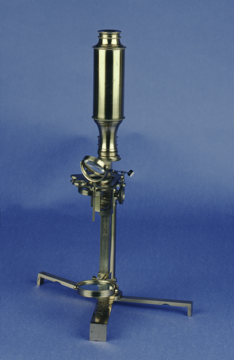
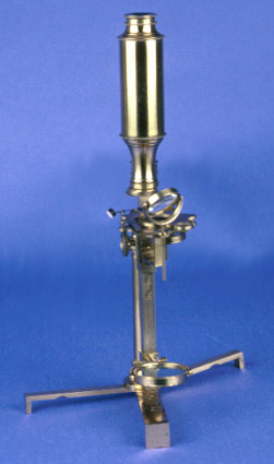
compound microscope, side pillar type, by Dollond, English, 1800 (c)
Origin
England; London
Maker
Dollond
Class
microscopes
Earliest Date
1800
Latest Date
1800
Inscription Date
Material
metal (brass, oxidised brass); glass; wood; ivory
Dimensions
overall height 443mm; breadth 240mm; depth 212mm box length 334mm; breadth 226mm; height 101mm
Special Collection
Robert Whipple collection
Provenance
Purchased from Bulles (?) & Co, Cambridge, on 29/12/1926.
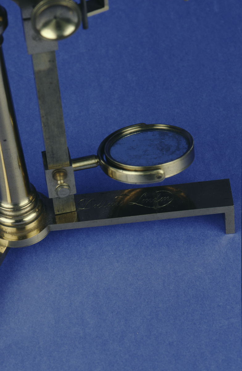
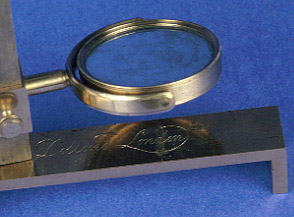
Inscription
‘Dollond London’ (foot)
Description Notes
Tribrach folding foot. Pillar with compass joint to limb. Limb with swinging plano-concave mirror on sliding shoe. Rack on limb to knurled screw on stage. Square stage with circular aperture and fittings for stage forceps etc. Arm pushes into slot at head of pillar and body screws to it. Snout with field lens pushes into collar with screw-fit eyepiece. Wheel of six objectives. Cone. Stage forceps with black and white ground. Substage condenser with swinging shoe (one screw missing). Stage condenser on rod. Frog plate. Live box with black and white ground. Lieberkuhn holder. Lieberkuhn holder for grounds, glasses etc.
Fitted wooden box.
Condition good; complete
References
Events
Description
The compound microscope was developed during the 17th Century and was closely related to the refracting telescope. Its popularity increased after the publication of Robert Hooke’s (1635-1703) Micrographia in 1665. Micrographia contained detailed pictures, never before seen, of insects magnified using a compound microscope.
A compound microscope uses two or more lenses. The lenses are held at certain distances from each other and are mounted inside a rigid tube. The tube was usually made from pasteboard, ivory, or most commonly, brass. The basic compound microscope magnifies an image in two stages -
Stage one: Light from a mirror is reflected up through the specimen into a powerful objective lens.
Stage two: The image produced by the objective lens is magnified again by the eye lens, which works like a simple magnifying lens.
The first compound microscope consisted of a simple barrel which would have been held up to the light. Later developments ensured that the compound microscope had a stable base, usually a brass stand and a side pillar.
In the 17th Century, the compound microscope had some serious drawbacks which made it easier to use a simple microscope (which have only one lens) instead. The image produced by a compound microscope was often affected by two types of aberrations known as chromatic and spherical. These aberrations caused blurring to the image (spherical) and the edge of the specimen to colour (chromatic). Chromatic aberration was removed at the end of the 18th Century by Harmanus van Deijlan, an instrument maker in Amsterdam. In 1830, spherical aberration was overcome by Joseph Lister, who developed the achromatic lens. Achromatic lenses became widely used in microscopes in the 1850s and are still used today.
FM:40074
Images (Click to view full size):